
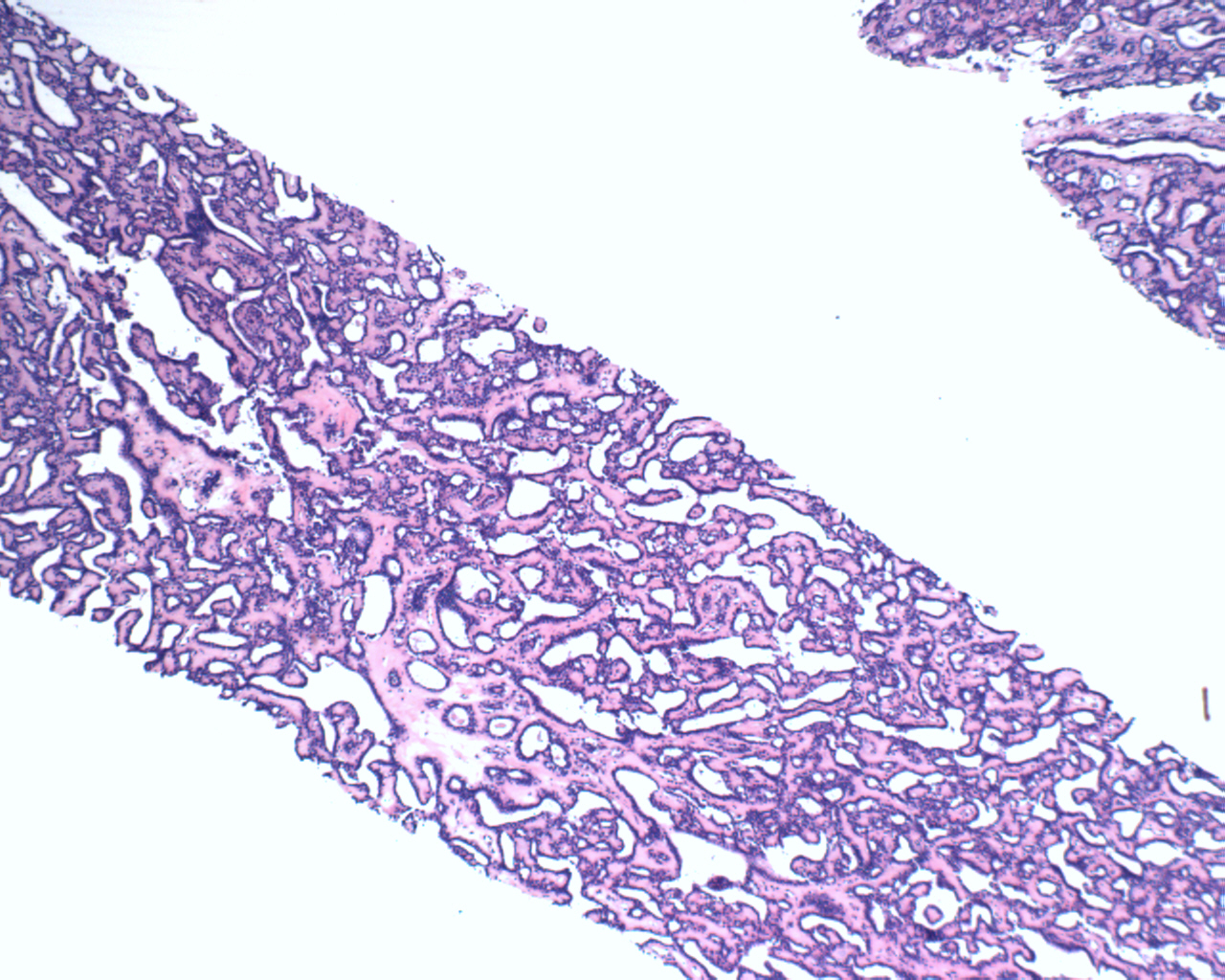
Subareolar biopsy demonstrates a proliferation of glands.
)
The ductal proliferations are lined by bland epithelial cells.
)
p63 and calponin combostain highlights the intact myoepithelial layer, confirming the benign nature of this lesion.
)
Nipple adenoma is not a specific term, but is used to characterize a subareolar lesion. This term most commonly describes a proliferation of glands arising from the nipple duct. Other terms that have been used for this lesion include subareolar papillomatosis or florid papillomatosis.
Histologically, the glandular structures proliferate and form either adenomatous or papillary structures. The epithelium is bilayered i.e. an intact myoepithelial layer is present. A pseudo-infiltrative pattern with entrapped tubules may be present at the periphery (Fletcher).
May occur anytime after puberty, but most patients are over 40. The lesion presents as an irregular firm mass and may become erythematous and crusted, mimicking Paget's disease of the nipple.
→May mimic Paget's disease of the nipple clinically.
Fletcher CDM, ed. Diagnostic Histopathology of Tumors. 3rd Ed. Philadelphia, PA: Elsevier; 2007: 916-7.