
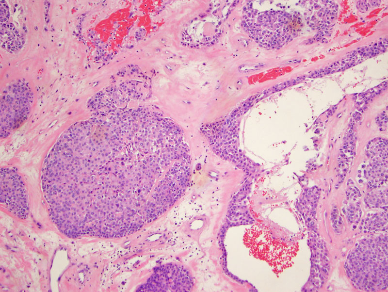
Solid nests and pseudocystic spaces with a eosinophilic hypocellular stroma; this arose as a well-demarcated nodule in the palm.
Well demarcated nests and a lobulated architecture are characteristic of this tumor. These tumors are usually well-circumscribed, but some do have focal infiltration into surrounding tissue. Most exhibit a lobulated architecture as seen here, and the tumor cells can be arranged in intersecting cords, sheets or nests. The stroma can be myxoid, chondromyxoid, collagenous or hyalinized.
The myoepithelial cells can be plasmacytoid.
Cytokeratin is strongly positive which is expected.
There was also focal EMA staining. A minority stain for SMA and GFAP and even calponon. S100 is usually positive, and desmin is negative.
Myoepithelial tumors are well-described in the salivary glands, but are also reported in the breast and soft tissues. Characterizing myoepithelial tumors in the soft tissues has been difficult due to their rarity. Furthermore, myoepithelial cells are very plastic and can assume a variety of morphologies (plasmacytoid, spindled, epithelioid, clear cell)(Hornick, Sasaguri).
Myoepthelial cells exhibit both epithelial and smooth muscle differentiation. In their study of 101 tumors, 93% were reactive for AE1/AE3, 87% for S100, 63% for EMA, 46% for GFAP, 36% for SMA, 23% for p63 and 14% for desmin (Hornick).
In the salivary gland, myoepithelial tumors that contain a ductular component are termed "mixed tumors or pleomorphic adenomas". Similarly in the soft tissue, a subset of myoepithelial tumors may also demonstrate ductular differentiation, and thus may also be classified as "mixed tumors". Nonetheless, this determination is clinically insignificant -- it is known that myoepithelial tumors and mixed tumors/pleomorphic adenomas exist on a spectrum.
A thornier issue is determining when a mixed tumor or myoepithelioma of the soft tissue becomes malignant. In the salivary gland, invasion beyond the tumor capsule is the main criterion for defining malignancy, whereas cytologic atypia and mitotic activity are supporting criteria. However, firm guidelines have not been established for these tumors in the soft tissue. Furthermore, many of these soft tissue tumors are unencapuslated, so the criterion of capsular invasion cannot be applied.
Hornick and Fletcher proposes that tumors with mild cytologic atypia be classified as myoepithelioma or mixed tumors whereas those with moderate or severe cytological atypia be classified as malignant myoepithelioma (myoepithelial carcinoma) or malignant mixed tumor.
Differential diagnosis for myoepithelioma of the soft tissue (especially if in the dermis) would include chondroid syringoma (sweat gland neoplasm), metastatic carcinoma, metastatic melanoma, epithelioid sarcoma and epithelioid leiomyosarcoma (Hornick, Sasguri).
In a review of 101 cases of myoepithelial tumors of soft tissue, Hornick et al (2003) found that that there were 53 males patients and 48 female patients (mean age 38, range 3-83). The tumors were concentrated in the extremities (41 in lower limbs, 35 in upper limbs), with the remainder in the head and neck (15 cases) and trunk (10 cases).
In general, the ones classifiable as myoepithelioma usually behave in a benign fashion despite their invasive nature. In one series containing 31 malignant cases, 42% recurred locally and 32% metastasized (lungs, lymph nodes). 4 patients died of their disease (Hornick and Fletcher).
• Salivary Gland : Myoepithelioma
• Myogenic : Leiomyosarcoma, Epithelioid Type
Hornick JL, Fletcher CD. Myoepithelial tumors of soft tissue: a clinicopathologic and immunohistochemical study of 101 cases with evaluation of prognostic parameters. Am J Surg Pathol. 2003 Sep;27(9):1183-96.
Sasaguri T, et al. Myoepithelioma of Soft Tissue. Pathology International 1999;49:571-576.

)
)
)
)
)