
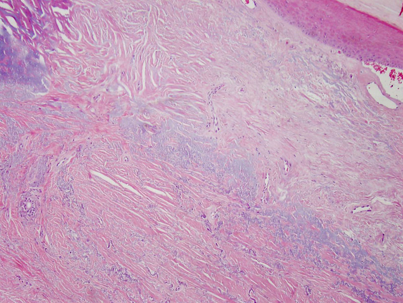
Low power view showing a small section of hyperkeratotic epidermis in the upper right corner and underlying dermis with amorphous basophilic elastotic material indicative of solar elastosis. One can appreciate the haphazard arrangement of collagen bundles toward the left side of the image and a slight basophilia to the dermis.
)
At higher power, once can appreciate elastic fibers dispersed between the collagen bundles.
)
The special stain for elastin better demonstrates the distribution of elastic fibers throughout the collagen bundles.
)
Burks was the first to describe this lesion in 1960 and coined the term ‘degenerative collagenous plaques of the hands’ based off five patients with a dermal alteration of collagen and elastin. Other terms used to describe this lesion include ‘keratoelastoidosis marginalis’, ‘collagenous and elastotic marginal plaques of the hands’, and ‘digital papular calcific elastosis’.
The exact etiology of this lesion has not been fully elucidated; Mehregan suggested that prolonged pressure and trauma in combination with ultraviolet radiation may be responsible.
Histologically, this entity is characterized by thick collagen bundles haphazardly arranged, some of the bundles are even oriented perpendicular to the epidermis. Amorphous basophilic material and elastic fibers can be seen in between the collagen bundles. The exact nature of this basophilic material is unclear, some authors believe it is degenerated elastic fibers or more likely, calcified deposits (Mortimore).
Collagenous and elastotic marginal plaques of the hands typically occurs in older men, with a reported mean age of 71 years (Jordaan). Individuals present with bilateral symmetric linear plaques that appear waxy with a white to yellow color. The lesions are located on the radial side of the hand at the junction of the dorsal and palmar skin.
Treatment is not generally necessary as individuals are usually asymptomatic.
Patients are rarely symptomatic as this is a slowly progressive process.
Burks JW, Wise LJ, Clark WH. Degenerative collagenous plaques of the hands. Arch Dermatol. 1960;82:362–6.
Jordaan HF, Rossouw DJ. Digital papular calcific elastosis: A histopathological, histochemical and ultrastructural study of 20 patients. J Cutan Pathol. 1990;17:358–70.
Mehregan AH. Degenerative collagenous plaques of the hands. Arch Dermatol. 1966;93:633.
Mortimore RJ, Conrad RJ. Collagenous and elastotic marginal plaques of the hands. Australas J Dermatol. 2001 Aug;42(3):211-3.
Weedon D. Weedon’s Skin Pathology. 3rd ed. Philadelphia, PA: Elvsevier; 2010:343-5.