
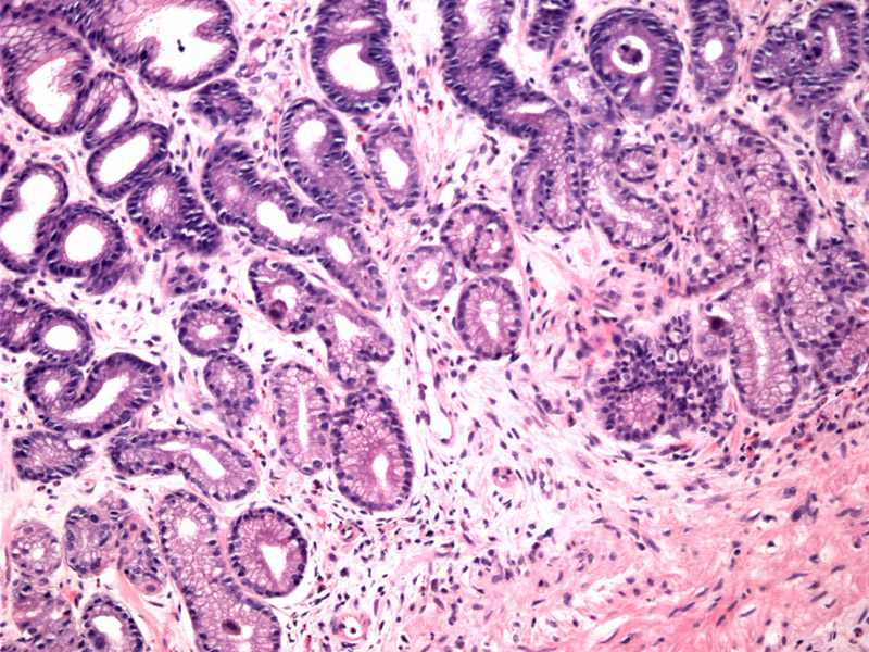
Viral inclusions in CMV gastritis are often present in the gastric glandular epithelium as well as endothelial cells and fibroblasts. At this magnification, however, such findings are not always easily confirmed
)
A closer look is necessary to identify the characteristic owl-eye inclusions characteristic of CMV. Note the enlarged epithelial cell within the mucus neck region.
)
Cytomegalovirus (CMV) most often infects the stomach and colon, within the GI tract. Enlarged cells with nuclear and cytoplasmic inclusions are seen. It is important to note that the inclusions in CMV gastritis are often present in the gastric glandular epithelium as well as mesenchymal cells (e.g. endothelial cells, fibroblasts). Inclusions are typically found deep within the ulcer base.
CMV gastritis is most commonly seen in immunocompromised individuals. Symptoms may include abbdominal pain, fever and weight loss. Endoscopically, one may see shallow or deep ulcers.
Ganciclovir or Vanganciclovir.
Case is contributed by Dr. Kate Sciandra, Dept of Pathology, VAMC Albuquerque NM.