
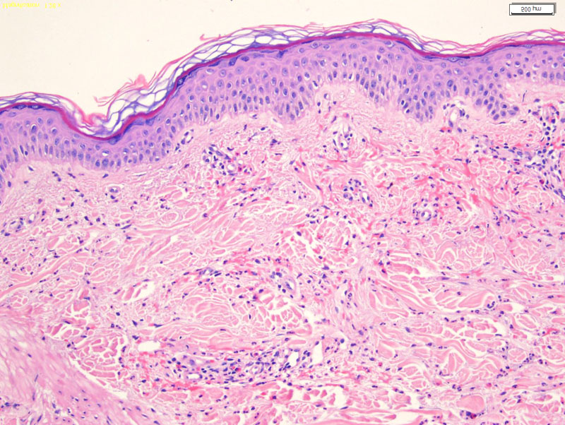
Urticaria is characterized by dermal edema, spare to moderate perivascular and interstitial inflammation composed of eosinophils, lymphocytes, neutrophils and mast cells (Rapini, Busam). The histology of urticaria is not very impressive. There is dermal edema and spare inflammation. The epidermis is usually normal.
)
A light inflammatory infiltrate is seen in a perivascular and interstitial distribution, composed of lymphocytes and many eosinophils. Neutrophils and mast cells may also be present.
)
Urticaria or hives is a common occurrence. Transitory and migrating wheals will move around different locations of the body for at least 24 hours. The wheals are very pruritic, and the process is mediated by type I hypersenstivity to allergens (foods, drugs).
Can be idiopathic or caused by drugs, infections, food allergies and as a reaction to cold (cold urticaria), heat or sun exposure.
Urticaria is usually self-limited and treated with antihistamines and avoidance of triggers (Busam).
- Mast cells are the primary effector cells
Busam KJ. Dermatopathology: Foundations in Diagnostic Pathology 1st Ed. Philadelphia, PA: Elsevier; 2010: 62-5.
Rapini RP.Practical Dermatopathology. Philadelphia, PA: Elsevier; 2005: 62.