
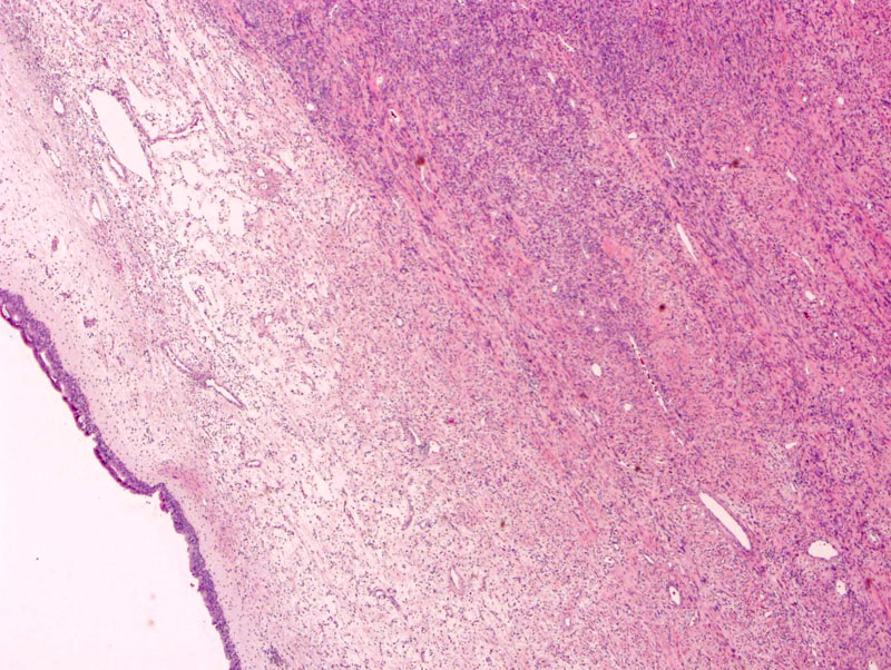
This SFT arose in the nasal cavity. Note the respiratory epithelium overlying the tumor (left image).
)
As with SFTs in other locations, it is composed of bland spindled fibroblasts admixed with ropey collagen.
)
The vasculature may show staghorn branching (not very obvious in this case). The spindle cells form a vague patternless pattern.
)
Solitary fibrous tumor is a mesenchymal tumor that can arise in in the pleura or a variety of extrapleural locations including the peritoneum, retroperitoneum, mediastinum, upper respiratory tract, orbit and soft tissues.
They are composed of bland, spindled CD34-positive fibroblasts in a "patternless" pattern with interspersed ropey keloidal collagen. The vasculature may demonstrate a staghorn, hemiangiopericytous pattern. The IHC profile of SFTs includ positivity for CD34, bcl-2 and CD99. The neoplastic cells are negative for S100, desmin or actin (Fletcher).
These are polypoid lesions can be excised in the sinonasal tract.
• Fibrous : Solitary Fibrous Tumor
• Pleura : Solitary Fibrous Tumor
Fletcher CDM, ed. Diagnostic Histopathology of Tumors. 3rd Ed. Philadelphia, PA: Elsevier; 2007: 92-3.