
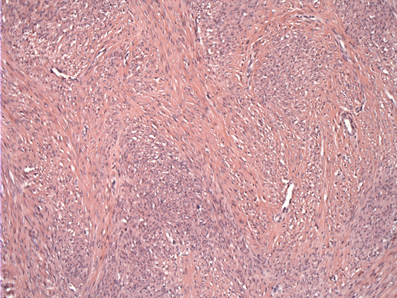
Fascicles of bland spindle cells, which are smooth muscle cells, will stain for progesterone and estrogen receptors. There are fusiform smooth muscle cells arranged in interdigitating fascicles with no noticeable atypia or mitotic figures.
)
Multiple rounded nodules with a uniform whorled appearance, morphologically similar to uterine myomas are seen studding the omentum, but can involve the peritoneum, in some cases the serosa of the uterus and fallopian tubes.
)
The spindled proliferation merges into fat. The cells are uniform and bland.
)
Leiomyomatosis Peritonealis Disseminata (LPD) is characterized by nodules of bland spindle smooth muscle cells studding the peritoneum. It is rare and about 100 cases have been reported in the literature (Summa). LPD is considered a metaplastic transformation of submesothelial pluripotent mesenchymal cells. The submesothelial location of the nodules as well as frequent findings of other metaplastic lesions (e.g. endometriosis, endoslpingiosis) support this theory (Fletcher).
Most arise in women of reproductive age and can occur in postmenopausal women. 40% of reported cases have occurred in black women. There is an association with pregnancy and oral contraceptive use, strongly indicating a hormonal role in pathogenesis (Fletcher).
When LPD occurs in pregnant women, there is often spontaneous regression after pregnancy. There have been about 10 cases of documented malignant transformation (Summa).
Fletcher CDM, ed. Diagnostic Histopathology of Tumors. 3rd Ed. Philadelphia, PA: Elsevier; 2007: 898-9.
Summa B et al. Leiomyomatosis peritonealis disseminata in a pregnant woman. Arch Gynecol Obstet. 2009 Apr 16. [Epub ahead of print]