
IMAGE DESCRIPTIONS
Case 1, image 1: This 31-year-old man died of multiple drug toxicity. He was in a moderate state of decomposition.
)
Case 1, image 2: This is what autolyzed lung looks like.
)
Case 1, image 3: It is hard to even make out the alveoli.
)
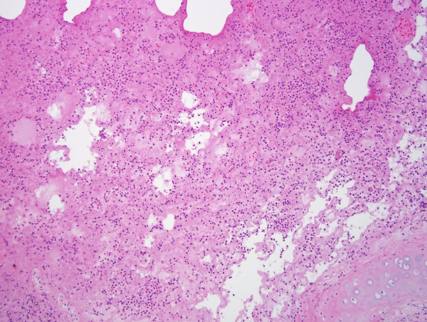
Case 1, image 4: The purple fuzzy stuff are bacterial colonies, probably postmortem overgrowth.
)
Case 1, image 5: If you search carefully, however, you will find better preserved areas with neutrophils within airspaces and thus, you can diagnosis early bronchopneumonia.
)
BACKGROUND
Despite significant postmortem changes (autolysis, putrefaction), taking sections for histology can still yield important information. For example, you can see steatosis in liver, fibrosis in myocardium and pneumonia in lung.
Last updated: 2013-10-22
For questions, comments or feedback on this case: editor@surgpath4u.com