
Intramuscular myxomas some infiltration into surrounding muscle fibers, splaying them apart. A few muscle fibers are seen here (eosinophilic islands) along the edge of th eleison.
)
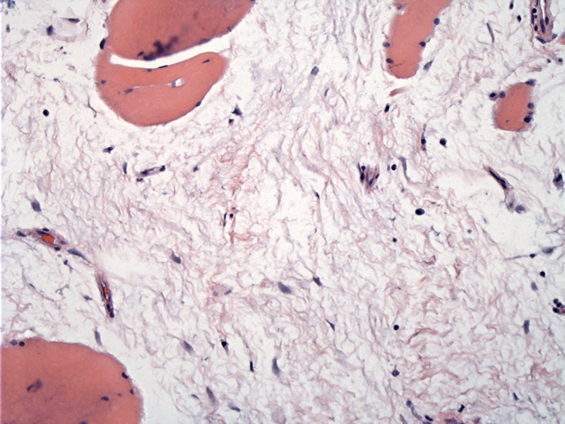
The tumor cells appear spindled or stellate with dark nuclei and are regularly but sparsely distributed in a copious loosely-textured mxyoid stroma. There are usually few blood vessels.
)
The stroma is myxoid and loosely textured. Blood vessels are largely absent or inconspicuous.
)
Myxomas are broadly divided into intramuscular (most common), juxta-articular or cutaneous mxyomas. Intramuscular myxomas mainly arise in muscle of thigh, upper arm and buttocks. Some patients develop multiple myxomas in association with fibrous dysplasia as part of the Mazabraud syndrome (Folpe, Fletcher).
Grossly, they appear gelatinous with cystic change and are fairly well-delineated from surrounding normal tissue.
Some myxomas are more cellular ("cellular myxomas") with a fascicular growth pattern with more pronounced vasculature, but still, areas of more typical myxoma should be present.
Differential diagnostic considerations include myxoid liposarcoma (exhibits an arborizing so-called chicken wire vasculature and the presence of atypical lipoblasts) and myxofibrosarcoma (again, prominent vasculature and the presence of pleomorphic tumor cells). S-100 can be helpful in differentiating myxoid neurofibroma or low-grade myxoid MPNST (positive in these tumors) and negative in myxomas. Intramuscular myxomas are paucicellular, hypovascular and bland.
Of interest, GNASI mutations seen in fibrous dysplasia have also been found in intramuscular mxyomas (Fletcher).
Occurs mostly in older adults with a female predilection. Presents as a slow-growing painless mass in the muscles of the thigh, buttocks or upper arm.
Benign; excision is curative.
Recurrence is extremely uncommon.
• Joints : Juxta-articular Myxoma
Fletcher CDM, ed. Diagnostic Histopathology of Tumors. 3rd Ed. Philadelphia, PA: Elsevier; 2007: PAGE.
Folpe AL, Inwards CY. Bone and Soft Tissue Pathology: Foundations in Diagnostic Pathology Philadelphia, PA: Elsevier; 2010: 276-7.