
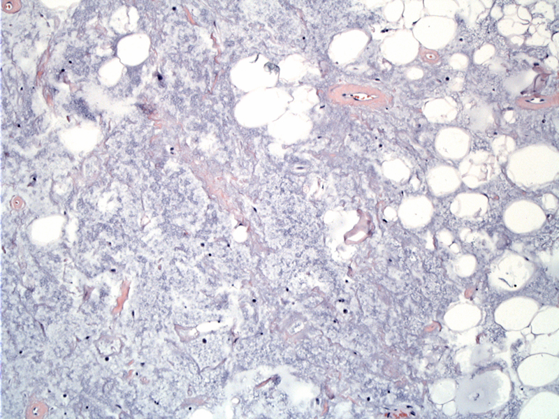
Part of this fatty tumor is replaced by zones of blue-grey mucoid substance. Also, note the interspersed strands of pink collagen in the stroma.
)
Ordinary fat is seen admixed with mucoid material. Small vessels are also apparent. Curvilinear capillaries are not seen.
)
Benign lipomas may occasionally show foci of myxoid degeneration and if that becomes extensive, the lesion is termed "myxolipoma". In some cases, the lesion exhibits increased vascularity and a prominent component of bland spindle cells and bundles of ropy collagen. These might be better described as myxoid variants of a spindle cell lipoma. Either way,it is simply important to recognize these benign variants as a myxoid spindle cell lipoma may occasionally elicit concern for a myxoid liposarcoma.
Occurs in adults and can arise anywhere in the body with the trunk and proximal limbs being the most common. Lipomas in the hands and feet are rare. Although both superficial and deep soft tissue lipomas exist, those arising in the abdomen or retroperitoneum should be thoroughly sampled as they are most likely well-differentiated liposarcomas (Fletcher).
Fletcher CDM, ed. Diagnostic Histopathology of Tumors. 3rd Ed. Philadelphia, PA: Elsevier; 2007: 1529.
Folpe AL, Inwards CY. Bone and Soft Tissue Pathology: Foundations in Diagnostic Pathology Philadelphia, PA: Elsevier; 2010: PAGE