
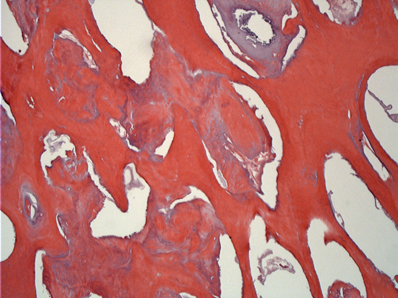
Case 1: Dentin or cementum-like matrix is in a haphazard arrangement, compatible with a complex odontoma.
)
Case 2: Mature dentin overlies predentin. The central pulp tissue was lost during processing. This orderly arrangement is compatible with a compound odontoma.
)
Fragments of pulpal tissue (spindle mesenchymal tissue) are associated with partially calcified matrix.
)
This axial CT scan demonstrates at hyperdense mass associated with the posterior maxillary dentition on the left side.
)
Odontomas are hamaratomas composed of odontogenic epithelium and mesenchyme. They are one of the most common odontogenic tumors. There are two types: compound and complex.
Compound odontomas recapitulate teeth and have organized architecture with the following components: mature dentin, enamel matrix (mature enamel is usually lost during processing), cementum, dentin pulp tissue, follicular tissue. Complex odontomas are composed of the same elements, except the arrangement is haphazard (Thompson).
Distinguishing between a developing complex odontoma and an ameloblastic fibro-odontoma may be impossible histologically (Barnes).
Most odontomas occur during the first two decades of life with an equal gender distribution. Most cases are asymptomatic and found incidentally on routine dental xrays. However, they can lead to swelling of the jaw with displacement of adjacent teeth as well as impaction of a permanent tooth.
Compound odontomas tend to arise in the anterior maxilla whereas complex odontomas are more likely to be in in the posterior mandible.
Radiographic: Tooth-shaped structures "toothlets" or radiodense masses are surrounded by a radiolucent zone (Thompson).
Surgical excision is curative.
→Odontomas are common odontogenic tumors and are hamartomas.
→They come into subtypes: complex and compound. Compound odontomas are "toothlets" and recapitulate teeth. Complex ondotomas are haphazardly arranged.
Barnes L, Eveson JW, Reichart P, Sidransky D. WHO Classification: Pathology and Genetics, Head and Neck Tumors Lyons, France: IARC; 2005: 310-1.
Thompson LDR, Wenig BM, eds. Diagnosis Pathology: Head and Neck. 1st Ed. Manitoba, Canada; Amirsys;2011; 6, 50-1.