
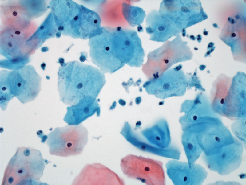
Pap smear with a few small Trichomonas organisms which appear lost among the background smaller cells.
)
Close inspection is required to definitively identify the blue gray organisms with tiny eccentric nuclei. The nucleus is necessary to identify Trichomonas, and to avoid misinterpretation of these organisms with mucus, degenerating neutrophils, or even metaplastic cells. The squamous cells show inflammatory type perinuclear halos.
)
An organism with granules (see arrow) is located in the center of the field. Perhaps the flagellum can be seen as well. Squamous cells show inflammatory hazy perinuclear halos.
)
Prominent granules are present within this organism. Arrow points to organism.
)
Inflammatory perinuclear halos involve many of the squamous cells. This finding should prompt a search for organisms as one possible etiology. Arrow points to the trichomonas.
)
Trichomonas vaginalis is a protozoal flagellated organism that is transmitted sexually. It is the most common non-bacterial, non-viral sexually transmitted infection and prevalence has been increasing in recent years.
Patients will present with a watery to foamy vaginal discharge and vaginal pain. The classic finding on exam is a "strawberry cervix." Trichomonas is typically diagnosed in the office via wet mount but more recently vaginal test kits are becoming available.
Trichomonas can be reliably treated with metronidazole. It is important when treating sexually transmitted diseases to ensure partner therapy as well.
There are few long-term sequellae to a trichomonas infection, though coinfection rates with other sexually transmitted organisms are high.