
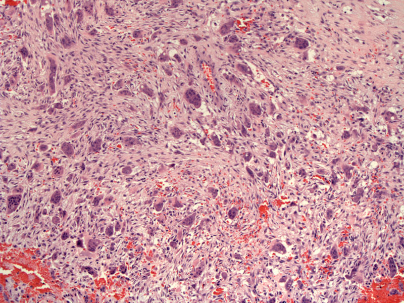
Peripheral giant cell reparative granuloma consists of a fibroblastic appearing background with interspersed red blood cells and multinucleated giant cells.
)
The giant cells have been shown to be highly similar to osteoclasts as are present in other giant cell lesions of bone.
)
Some areas may appear more spindled and devoid of giant cells.
)
Mature woven or lamellar bone is present in up to 1/3 of cases.
)
The bone is lined by osteoblasts and there is no atypia present.
)
All age groups were found to be affected with a relative predilection from 40 to 60 years of age. Peripheral giant cell reparative granuloma is an uncommon but not rare exophytic lesion of the oral cavity. The mandible is more often affected than the maxilla. Clinically, the lesions present as red smooth and rubbery or with a soft consistency.
Excision
Excellent. May recur if incompletely excised.