
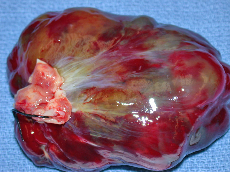
A globoid thinly encased mass attached to the endomyocardium comprises this atrial myxoma. There is extensive hemorrhage.
Hemorrhage is universally found within a myxoma. Although the tumor is dissected by hemorrhage, one can still pick out the myxomatous matrix (blue and frothy) and the myxoma cells within the matrix. Hemorrhage is thought to be a direct consequence of the mechanical trauma due to this tumor's mobility.2
This image demonstrates a more diagnostic area of the tumor with stellate myxoma cells in a mxyomatous background. Mitoses are not present this tumor and if found, another diagnosis must be considered.
These striking purple spikes are called Gamma-Gandy bodies, which are connective tissue fibers with iron and calcium deposits. Gamma-Gandy bodies are a consequence of past episodes of hemorrhage.
Again, this image demonstrates evidence of past hemorrhage with extensive Gamma-Gandy bodies and hemosiderin-laden macrophages.
Longer standing myxomas develop significant fibrosis. If significant, the underlying nature of the lesion can be difficult to identify.
Dilated glands are seen in clusters within this other case of atrial myxoma. Many glands show an attenuated lining, but other glands are lined by cuboidal or columnar cells (as seen below). This finding of glandular structures although characteristic of atrial myxomas, is seen in less than 5% of cases. Note the more typical area (upper right corner) consisting of singular myxoma cells dispersed in a background of myxoid stroma.
Other glands are lined by columnar cells with well-developed apical mucin.
The majority of tumors found in the heart are metastatic and primary tumors are fairly rare. Of the primary tumors of heart, myxomas are by far the most common. Although these tumors can occur in any of the four chambers including heart valves, they have a definite predilection for the atria, especially in the left atria near the fossa ovalis. Hence the commonly used term atrial myxomas.
Grossly, the mass can be sessile or pedunculated (connected to the atrial wall by a fibrous stalk). In its pedunculated form, the tumor can bob up and down through the AV valves during ventricular contractions and cause periodic obstruction. The tumor can also bounce against the valves, causing mechanical damage.1
Cardiac mxyomas are composed of a cellular component set against a myxomatous background. The cellular component consists of several types of lesional cells, all of which are thought to be derived from a primitive multipotent mesenchymal stem cell. The most prominent tumor cell is the mxyoma cell, also known as the lepidic (Gr: scales) cell because polyheydral mxyoma cells can resemble scales.2 The mxyoma cells can also have a fusiform or stellate morphology.
Other cell types of this tumor include spindle-shaped cells (smooth muscle differentiation), endothelial cells lining abnormal vascular structures (endothelial differentiation) and columnar cells forming a gland (glandular differentiation). Note that tall columnar cells that secrete mucin lining a gland or a poorly formed vessel is an distinctive characteristic of this tumor.
Palpitation, fever, and weight loss are the most frequent symptoms of cardiac myxomas, but cerebrovascular accident or peripheral arterial embolism are also seen. Systemic embolism is encountered in 30%-45% of cases . In individuals over 75 years of age, myxomas are most often nonobstructive, well-circumscribed, calcified tumors, with a low risk of obstruction or embolism. It has been suggested that only independent predictor of systemic embolism in left atrial myxoma is the polypoid nature of the lesion.
Mxyomas may occur in younger patients with a familial autosomal dominant syndrome called Carney's syndrome (aka Myxoma syndrome or Swiss syndrome). Cardiac myxomas from these patients have an increased tendency to be multicentric and recur more often after excision (21% recurrence in Carney's syndrome vs. 1% recurrence in sporadic lesions)2
Carney's syndrome, also known as "familial endocrine myxolentiginosis", has a complex manifestation, but it will be enough to remember the following lesions: (1) multiple cardiac and extra-cardiac (especially cutaneous) myxomas, (2) spotty skin pigmentation (especially blue nevi) and (3) endocrine overactivity (pituitary tumors or microadneomas of the adrenal gland).
Surgery is the treatment of choice. The superior transeptal approach has been reported to be an excellent approach with a nice surgical view to expose and remove a left atrial myxoma.
Benign. Rarely, the tumor can recur and thus, excision with a wide margin is recommended. Another rare complication is embolization (ie. from the left atria to the internal carotid artery).
→Key histologic features include: (1) fusiform or stellate cells in a myxoid background (2) hemorrhage (3) Gamma-Gandy bodies (4) Glandular structures lined by mucinous cells.
1 Kumar V, Abbas AK, Fausto N. Robbins and Cotran Pathologic Basis of Disease. 7th Ed. Philadelphia, PA: Elsevier; 2005: 613-4.
2 Fletcher CDM, ed. Diagnostic Histopathology of Tumors. 3rd Ed. Philadelphia, PA: Elsevier; 2007: 9-15
Carney JA, Hruska LS, Beauchamp GD, Gordon H. Dominant inheritance of the complex of myxomas, spotty pigmentation, and endocrine overactivity. Mayo Clin Proc 1986;61:165–72
Bire F, Roudaut R, Chevalier JM, Quiniou G, Dubecq S, Marazanoff M, et al. Cardiac myxoma in patients over 75 years of age. Report of 19 cases [French]. Arch Mal Coeur Vaiss 1999;92:323–8
Ha JW, Kang WC, Chung N, Chang BC, Rim SJ, Kwon JW, et al. Echocardiographic and morphologic characteristics of left atrial myxoma and their relation to systemic embolism. Am J Cardiol 1999;83:1579–82
Kunitomo R, Okamoto K, Utoh J, Nishimura K, Muranaka T, Tsurusaki S, et al. Evaluation of superior transseptal approach for the removal of left atrial myxoma [Japanese]. Kyobu Geka 2001;54:211–4

)
)
)
)
)
)
)
)