
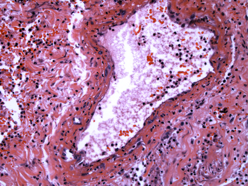
The venous wall is infiltrated by acute and chronic inflammatory cells.
)
The vessel wall (lower) is infiltrated by many inflammatory cells, and surrounds an organizing luminal thrombus (upper). The nodule in the center is an area of organizing fibrosis.
)
Thrombophlebitis is defined as inflammation of veins due to the formation of a thrombus. If inflammation is not present, the term phlebothrombosis may be used. However, in reality, the two entities cannot be definitively separated as they are spectrums of the same disease process. Remember the Virchow's triad? Stasis, endothelial injury and hypercoagulability lead to thrombus formation.
In superficial thrombophlebitis, the superficial veins are involved (such as the saphenous vein) and the vein may be tender with inflammation of the overlying skin. There is usually little edema involved in the affected extremity, and this condition is usually not life-threatening. However, in certain instances, the thrombus may extend into the deep veins, leading to deep vein thrombosis and potentially pulmonary embolus. Therefore, it is important to thoroughly evaluated patients with superfical vein thrombosis.
Deep vein thrombosis often presents with swelling of the lower extremity and tenderness of the vein. Diagnostic tests include D-dimer levels, duplex ultrasonography and MRI.
1 Rosai, J. Rosai and Ackerman's Surgical Pathology. 9th Ed. Philadelphia, PA: Elsevier; 2004: 2455.