
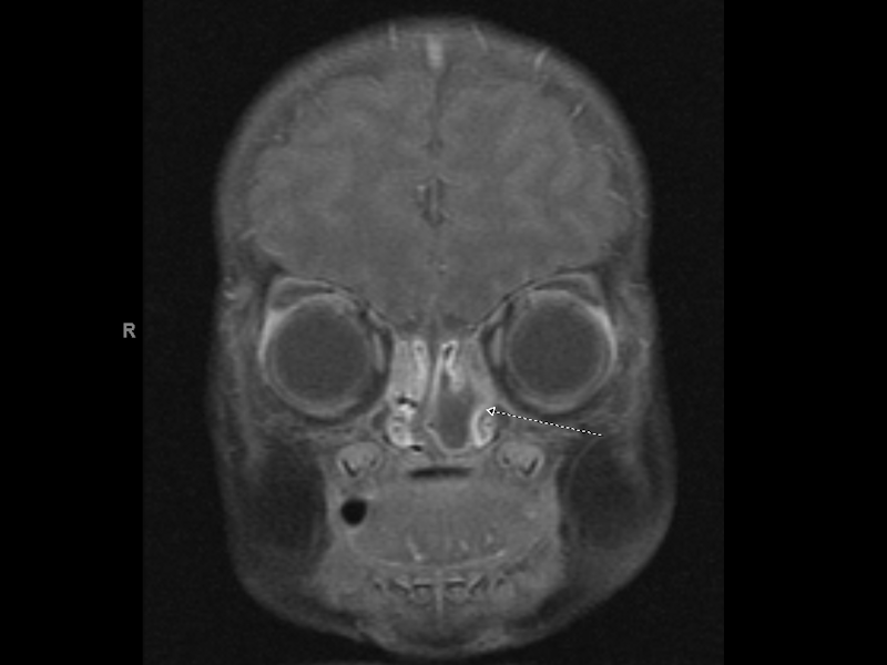
There is a T1 hypointense, T2 hyperintense rim-enhancing mass within the left nasal passage. The internal structure of the mass is primarily cystic but does appear to contain septae. There is no intracranial or intraorbital extension. The cribriform plate appears grossly intact. The mass causes left-to-right nasal septal deviation.
)
The overlying respiratory mucosa is intact, and the lesion undermines the epithelium.
)
A higher power view shows mucous glands of the nasal mucosa in a background of glial tissue and fibrosis.
)
Astrocytic cells with gemistocytic change (reactive astrocyte that is plump with lots of pink cytoplasm)are scattered within the glial tissue.
)
In areas such as this, the glial tissue could be easily overlooked at first glance. High power such as in this image, however, clearly reveals the diagnosis.
)
Almost all are present at birth, but rare lesions have been found in patients up to age 50. 1.5 to 3x more predominant in males. 25% occur posterior to nasal bone and form intranasal polypoid masses (presents with nasal obstruction and respiratory distress), 60% occur anterior to nasal bone and form subcutaneous lesions (firm round nodules on the bridge or side of the nose, can get quite big). Other rarer locations are nasopharynx, palate, paransala sinuses, tonsillar and pterygonpalatine fossa.
Cured by excision
Benign, rare local recurrences.
(1) AFIP
(2) Foundations in Diagnostic Pathology