
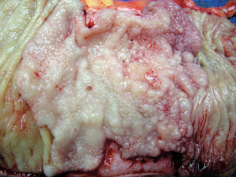
A pale glistening right sided mass suggests the high content of mucin. Note that normal bowel is seen on the right and left edge of the image. This morphology, which includes its exophytic and well-delineated margins, is compatible with an HNPCC related adenocarcinoma.
)
Glistening, almost gelatinous-appearing nodules stud the omentum in a patient with metastatic mucinous adenocarcinoma of the colon.
)
A peritoneal implant exhibits invasion by mucinous signet ring cells.
)
Ascites fluid shows single signet ring cells in a background of mesothelial cells and lymphocytes. The malignant cells have darked chromatin and an eccentric nucleus.
)
Another case shows a transition from conventional adenocarcinoma (left), to a poorly-differentiated signet ring cell type (right). This case proved to be MSI related. Interestingly, if the signet ring cell component was only a minor area, in the current practice of pathology, there is no definitive rule on how to report this finding. One reasonable approach is for pathologists to report the percentage of signet-ring cell component in colorectal mucinous adenocarcinomas (Sung).
 adjacent to mucinous areas. ', 'micro', ''))
The cellular areas show signet ring cells, which contain a belly of intracytoplasmic mucin that pushes the nuclei to the periphery.
)
Another area of signet ring cells floating in pools of mucin.
)
Some areas contain acellular mucin (mucin without the presence of malignant cells).
)
The junction of a cellular area to acellular mucin pools may appear abrupt.
)
Mucinous (colloid) adenocarcinomas comprise 10% of all colon cancers and up to 30% of rectal cancers. They are more common in patients with HNPCC (hereditary nonpolyposis colorectal carcinoma) and in carcinomas arising from a sessile serated adenoma -- in other words, there is an association with the microsatelite instability pathway of carcinogenesis.1 Many conventional-type colorectal carcinomas produce mucin, however, when more than 50% of the tumor is mucinous, the tumor can be classified as the mucinous subtype.1
Mucinous carcinomas have malignant glands 'swimming' in copious amounts of extracellular mucin. The mucin can be seen dissecting through the bowel wall. A variant of mucinous adenocarcinoma is signet ring adenocarcinoma, in which the mucin is intracellular rather than extracellular. Like its moniker, signet ring cells have a large belly of cytoplasmic mucin that pushes its nuclei to the periphery. If poorly-differentiated, the malignant signet ring cells may not have a signet-ring configuration, but simply resemble pleomorphic cells containing intracytoplasmic mucin. When signet ring cells comprise over 50% of the mucinous tumor, the tumor can be designated as a signet ring adenocarcinoma.1,2 . It is important to note that in direct comparison with signet ring adenocarcinoma, the mucinous type frequently presents with a lower TNM stage and with vascular invasion and lymph node involvement.
Ogino and colleagues (2006) conducted molecular profiling on 39 signet ring carcinomas, 167 mucinous carcinomas and 457 nonmucinous colorectal carcinomas -- demonstrating that signet ring carcinomas and mucinous carcinomas are related subtypes of colorectal carcinomas, but have molecular features that are also distinct from each other. For example, signet ring and mucinous carcinomas are more likely to have BRAF mutations, MSI and loss of MLH1 compared to nonmucinous carcinomas. Furthermore, mucinous carcinomas are more likely to have KRAS mutations and TP53 positivity and signet ring cells are less likely to have 18q LOH and COX2 overexpression.
Signet ring cell carcinomas have a poor prognosis and it is important to exclude metastasis from a gastric primary lesion.1,2 Colorectal signet-ring cell carcinoma has adverse prognostic significance independent of the stage at presentation (Kang). Mucinous histology is an independent adverse prognostic factor in some studies, but not in all.(Consorti; Halvorsen)
Signet ring cell carcinomas that grow in an exophytic and well-circumscribed manner are associated with HNPCC and sporadic colorectal cancers with DNA microsatellite instability. These have a more favorable prognosis. Undifferentiated carcinoma is also associated with DNA MSI and again also have a fair prognosis. Thus, it is interesting to note that tumors with DNA MSI, despite having unfavorable histology, carry a relatively fair prognosis.1,2
• Colon : Adenocarcinoma, Conventional Type
• Colon : Adenocarcinoma, Mismatch Repair Related
• Colon : Mucinous Adenocarcinoma
• Colon : Adenocarcinoma, Conventional Type
1 Iacobuzio-Donahue CA, Montgomery EA. Gastrointestinal and Liver Pathology: Foundations in Diagnostic Pathology. Philadelphia, PA: Elsevier; 2005: 382-9.
2 Fletcher CDM, ed. Diagnostic Histopathology of Tumors. 3rd Ed. Philadelphia, PA: Elsevier; 2007: 401.
3 Ogino S, Brahmandam M, Cantor M et al. Distinct molecular features of colorectal carcinoma with signet ring cell component and colorectal carcinoma with mucinous component. Mod Pathol. 2006 Jan;19(1):59-68.
Kang H, O'Connell JB, Maggard MA, et al. A 10-year outcomes evaluation of mucinous and signet-ring cell carcinoma of the colon and rectum. Dis Colon Rectum 2005;48:1161–1168. |
Consorti F, Lorenzotti A, Midiri G, et al. Prognostic significance of mucinous carcinoma of colon and rectum: a prospective case–control study. J Surg Oncol 2000;73:70–74.
Halvorsen TB, Seim E. Influence of mucinous component on survival in colorectal adenocarcinomas: a multivariate analysis. J Clin Pathol 1988;41:1068–1072.
Sung CO, Seo JW, Kim KM, Do IG, Kim SW, Park CK. Clinical significance of signet-ring cells in colorectal mucinous adenocarcinoma.Mod Pathol. 2008 Dec;21(12):1533-41.
***Gross images courtesy of Myra Zucker, Dept of Pathology at University of New Mexico, Albuquerque NM.