
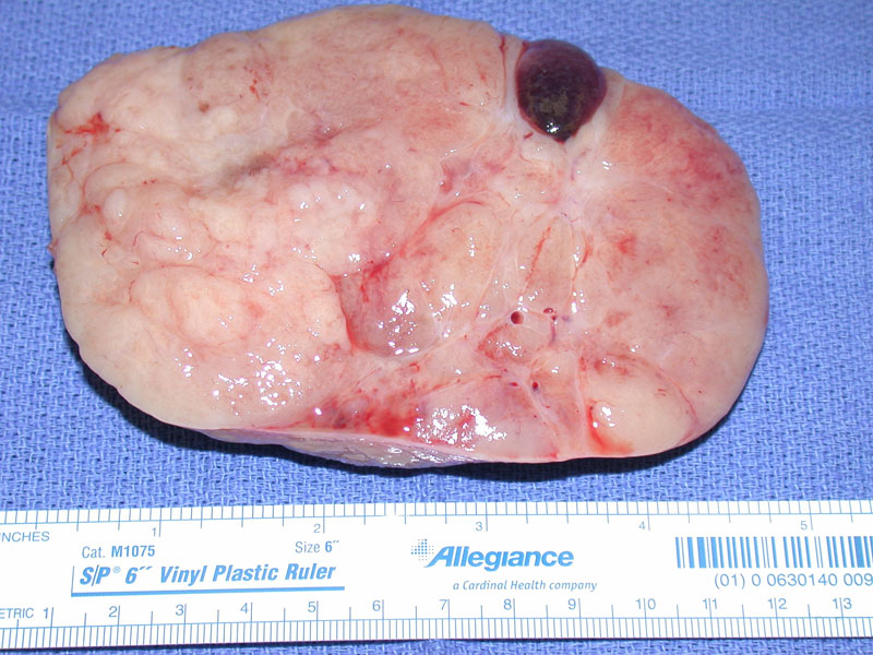
This cut surface of a resected dysgerminoma is typical, showing a solid uniform pale tan appearance. A focus of hemorrhage is also seen. No cystic areas are seen to suggest a teratoma component.
.', 'gross', ''))
Dysgerminoma is comprised of a uniform population of polygonal cells with distinct cell membranes, abundant clear to granular cytoplasm. The nuclei are centrally or eccentrically located with
)
The tumor cells are arranged in nests and cords, separated by a fibrous stroma containing lymphocytes. The lymphocytes are usually confined to the fibrous stroma, but occasionally may be seen amongst the tumor cells.
)
Poorly circumscribed nests of tumor cells admixed with lymphocytes are set against a fibrous stroma. The tumor cells here contain more granular cytoplasm, rather than the classic clear cytoplasm.
)
Dysgerminomas characteristically stain positively with PLAP, CD117 and OCT4. In this image, the tumor cells have strong cytoplasmic staining with PLAP (placental alkaline phosphatase). OCT4 is a nuclear transcription factor, and thus, would demonstrate nuclear staining.
)
This area of a different tumor shows a preponderance of background lymphocytes, which separate the malignant cell population.
)
A few lone ovarian follicles hold out among the background malignant cells.
)
A fairly distinct granuloma is evidenced here as a collection of epithelioid histiocytes with pink cytoplasm and a small rim of lymphocytes.
)
This latter case was associated clinically with elevated HCG but no synctiotrophoblast appearing cells were found microscopically. The immunostain for HCG does highlight scattered cells which morphologically appear as the usual dysgerminoma type cell.
)
Dysgerminoma is the ovarian counterpart of the testicular seminoma. It is the most common malignant germ cell tumor of the ovary, and the most common gonadal tumor in the setting of gonadal dysgenesis.1
Grossly, the tumor is solid and large (at least 10cm in diameter). The cut surface is tan, fleshy and lobulated. Microscopically, ovarian dysgerminoma is virtually identical to the testicular seminoma. The tumor cells are monotonous, polygonal cells with abundant clear cytoplasm (resembling primordial germ cells). They frequently grow in islands and sheets, separated by a fibrous stroma containing lymphocytes. The lymphocytic component can form germinal centers and the infiltrate can be so prominent that it obscures the tumor cells.2
Dysgerminoma is a tumor of young women -- 90% of patients are under 30 years old (average age of diagnosis is 22). Twenty percent of cases are detected during pregnancy. Typically presents with a rapidly enlarging abdominal mass accompanied by abdominal distention and pain. Serum LDH is always elevated and serves as a useful tumor marker. Placental alkaline phosphatase (PLAP) is usually elevated as well, and some clinicians use both LDH and PLAP to monitor the progression of the tumor.1
Increased levels of alpha-fetoprotein (AFP) or human chorionic gonadotrophic (HCG) implies the presence of other germ cell elements such as yolk sac component (AFP) and trophoblastic disease (HCG). Approximately 3% of dysgerminomas contain syncytiotrophoblastic giant cells, which stain positively with HCG, and is clinically detected by a low level of HCG.2
60-80% of patients present at stage I (confined to the ovaries). The tumor is usually unilateral, but may be bilateral (stage Ib) in 5-15% of cases. For patients who have unilateral disease wishing to preserve fertility, unilateral salpingo-oophorectomy may be performed. However, biopsy of the contralateral ovary is recommended as the tumor may be microscopic. If patients are not interested in preserving fertility, full staging is performed with total hysterectomy, bilateral salpingo-oophorectomy, pelvic lymphadenectomy, omentectomy, washings, and peritoneal biopsies of the pelvis, abdomen, and diaphragm. The standard chemotherapy regimen for germ cell tumors of the ovary is BEP (bleomycin, etoposide, cisplatin).
If the tumor is truly confined to one ovary (stage Ia), overall 5 year survival is over 90%. Even if the tumor has spread beyond the ovary, long-term prognosis is still good as the tumor is exquisitely sensitive to platinum-based chemoreagents. Overall 5-year survival is between 75-90%.1,2
♣ Dysgerminoma is the ovarian counterpart of the testicular seminoma. Histologically, they are identical.
♣ Serum levels of LDH and PLAP are often helpful to track the progression of the tumor. Modest elevations of AFP suggest a component of syncytiotrophblastic giant cells.
♣ Dysgerminoma is a tumor of young women, with average age of diagnosis at 22.
• Testis : Seminoma, Classic Type
1 Nucci MR, Oliva Esther. Gynecologic Pathology: Foundations in Diagnostic Pathology. Philadelphia, PA: Elsevier: 2009: 501-7.
2 Fletcher CDM, ed. Diagnostic Histopathology of Tumors. 3rd Ed. Philadelphia, PA: Elsevier; 2007: 604-5.
3 Kumar V, Abbas AK, Fausto N. Robbins and Cotran Pathologic Basis of Disease. 7th Ed. Philadelphia, PA: Elsevier; 2005: 1101.