
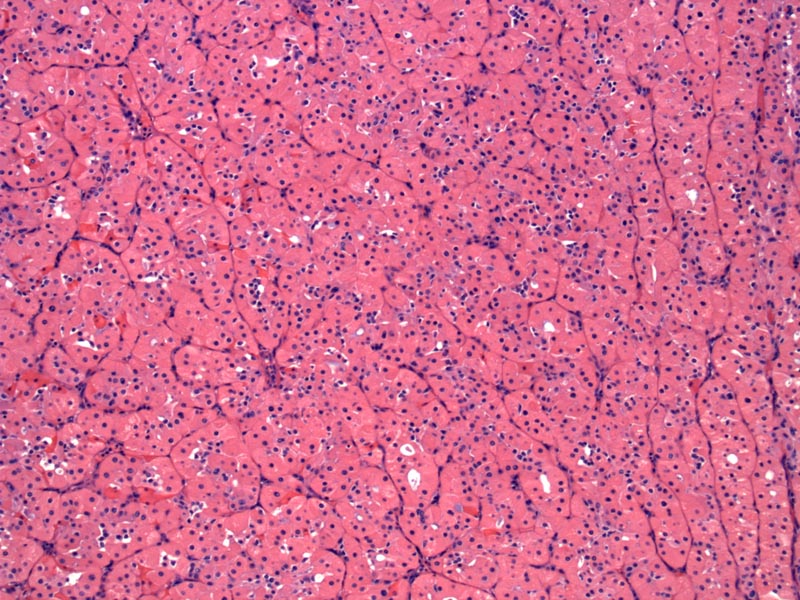
The tumor is composed of solid sheets or nests of oncocytes, which have abundant eosinophilic cytoplasm and uniform round nuclei.
)
A higher power view shows oncocytes with centrally placed nuclei, some with prominent nucleoli. This is a benign tumor, whereas oncocytic carcinoma would demonstrate significant pleomorphism and necrosis.
)
The gross appearance is a well-circumscribed, mahogany brown tumor with a uniform consistency.
)
FNA with Diff-Quik: Oncocytes on have blue waxy cytoplasm.
)
FNA with Pap stain: The oncocytes have a more granular pink cytoplasm with this stain. The cells are uniform with a round nucleus.
)
Oncocytomas are rare benign salivary gland tumors, thought to be dervied from striated cells of the salivary gland (which are mitochondria rich). They are quite rare (1-3% of salivary gland tumors).
Oncocytes have a polygonal shape, with abundant eosinophilic cytoplasmic (packed with abnormal mitochrondria) and centrally placed nuclei (with or without prominent nucleoli). Sometimes, oncocytes may accumulate glycogen and have a clear cell appearance (not shown here); if that is the predominant pattern, then it is called a clear-cell oncocytoma.
On cytology, aspirated oncocytes are indistinguishable from oncocytes of a Warthin tumor. Therefore, one needs to look at the background. Warthin tumor has abundant lymphocytes and relatively few oncocytes and vice versa for oncocytoma. Other diagnostic considerations include oncocytic metaplasia and oncocytosis. However, as all the aforementioned entities are benign, it is not absolutely crucial to make a distinction.
In some aspirates, on low power, it may be difficult to distinguish the cells from an oncocytoma from tumors cell in acinic cell carcinoma (in which cells can have dense cytoplasm and round uniform nuclei). Furthermore, both oncocytoma and ACC may have clear cell change. Upon closer inspection, the cytoplasm in ACC cells have vacuolated cytoplasm. IHC may help as oncocytes will stain with PTAH (phosphotungstic acide hematoxylin) and anti-mitochondrial antibodies. The cytoplasmic granules in ACC cells will be PAS-positive and diastase-resistant (Cibas).
Usually occurs in the 6th to 8th decade of life, affecting both genders equally. Most common location is the parotid gland (~85%), with the submandibular gland the next most common site. Presents as a slow growing mass. Approximately 20% of patients have a history of radiation exposure (Fletcher).
Cibas ES, Ducatman BS Cytology:Diagnostic Principles and Clinical Correlates 3rd Ed. Philadelphia, PA: Elvesier; 2009: 302-3.
Fletcher CDM, ed. Diagnostic Histopathology of Tumors. 3rd Ed. Philadelphia, PA: Elsevier; 2007: 262-5.