
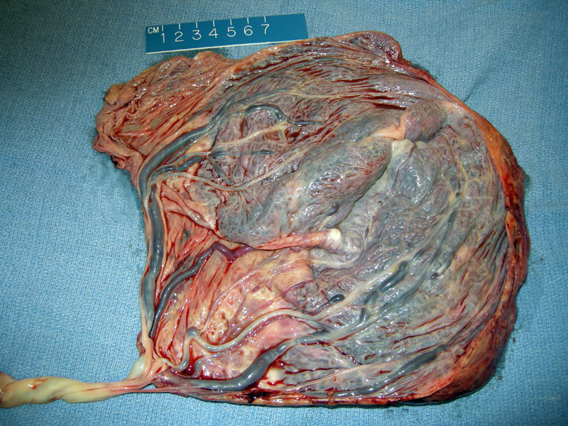
Bilobed placenta with velamentous cord insertion is seen.
)
The umbilical cord normally inserts near the center of the placenta. In 7% of term singleton placentas, the cord inserts at the edge of the placenta, called 'marginal insertion'. In 1% of cases, the cord inserts even further away from the placenta (in the membranes) and thus is called 'velamentous or membranous insertion'.
Velamentous cord insertions are more common in twin births, when they course in the dividing membranes of the twin placenta. There is also an association with single umbilical artery.
When the umbilical vessels ramify through the free membranes, they are no longer protected by Wharton's jelly and thus become vulnerable to trauma and shearing forces. If these membranous vessels pass over the internal cervical os, this is called 'vasa previa' and these vessels are easily ruptured during vaginal delivery. 1 In addition to the risk for vasa previa, these vessels are more likely to be compressed during labor resulting in fetal heart rate decelerations. There is also an association with intrauterine growth restriction.
In the absence of vasa previa, no treatment is necessary. In cases of vasa previa, a cesarean delivery is necessary to avoid trauma to fetal vessels.
While risks of intrauterine growth restriction, nonreassuring surveillance, and vasa previa exist, most cases are uncomplicated and the velamentous insertion is an incidental finding at delivery.
1 Baergen RN. Manual of Benirschke and Kaufmann's Plathology of the Human Placenta. New York, NY: Springer; 2005: 263-6.