
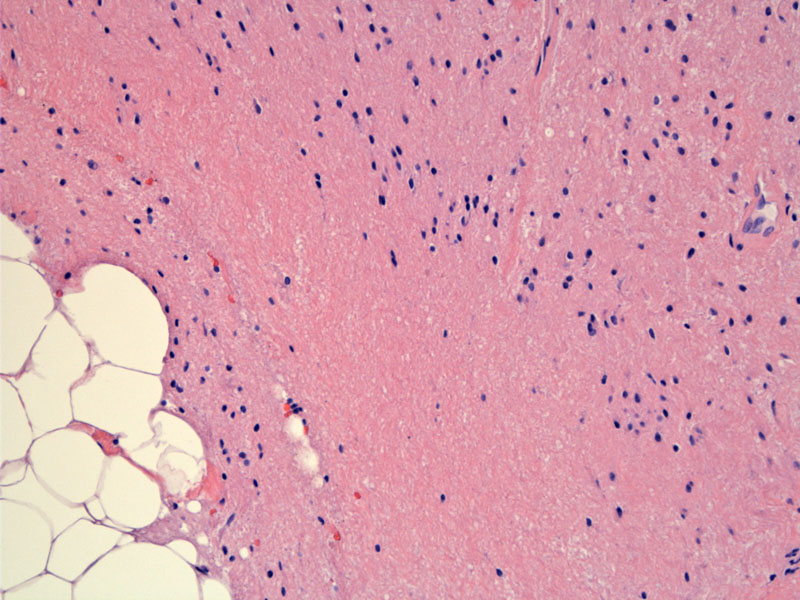
Mature glial tissue (right) abuts mature adipose tissue (left).
)
Glial tissue underlies ciliated columnar epithelium (respiratory epithelium) typical of the nasopharynx.
)
Squamous-lined cyst with adnexal structures (sebaceous glands, hair follicles) are the most common findings in mature cystic teratomas.
)
A disrupted piece of hair (cylindrical brown pink bodies with vertical stripes) is surrounded by foreign body giant cells and keratin debris. Disrupted cystic contents often elicit a prominent foreign body granulomatous response.
)
Mature glial tissue (upper left), fat (center), and cartilage (bottom right) can be appreciated in this image.
, fat and cartilege (bottom right) can be appreciated.', 'micro', ''))
Osteoid may also be encountered.
)
Mature cystic teratomas are the most common ovarian neoplasm (25% of all ovarian tumors) and accounts for the vast majority of germ cell tumors. These tumors are thought to arise from parthenogenesis -- either from fusion of the ovum with the second polar body or suppression of meiosis II (second reductional division of chromosomes). In Greek, parthenos means 'virgin' and genesis means 'creation', therefore, parthenogenesis = virgin birth or asexual reproduction. For those of you who remember Greek mythology, Athena Parthenos was the Goddess of Wisdom who sprung from Zeus' head.
Grossly, these tumors are less than 10 cm in diameter with a smooth external surface. The cut surface is cystic and may contain abundant hair, with oily and sebaceous material. A white nodule containing bone or teeth may protrude into the cystic lumen, the so-called Rokitansky protuberance. In very rare instances, a structure resembling a malformed fetus (homunculus) may be seen attached to the cystic wall. Dense solid areas are uncommon and should raise the possibility of an immature teratoma. 10-15% of tumors are bilateral.1,2
Microscopically, the tumor is composed of mature tissues from one or more embryonic germ cell layers. Two-thirds of teratomas contain two germ layers and one-third contain all three germ layers. Ectodermal derviatives (skin, hair appendages, sebaceous and sweat glands) are the most common, and when they comprise a dominant portion of the neoplasm, the term 'dermoid cyst' is commonly employed. Other ectodermal elements include brain (glia), choroid plexus, peripheral nerve and dental structures.1,2
Endodermal elements include gastrointestinal and respiratory mucosa, thyroid tissue and renal tissue. Mesodermal derivatives include fat, muscle, bone, cartilage and loose connective tissue. Cytologic atypia and mitotic activity are largely absent. A prominent foreign body granulomatous response due to rupture cyst contents is quite common.1,2
Wide age range is seen with 50% of cases occur between ages 20-40 (peak incidence between 20-29 years). Half the patients are asymptomatic (discovered incidentally during evaluation or treatment for another condition), and the other half may present with abdominal pain and abnormal uterine bleeding. Torsion and rupture are the most serious complications, which occur in 1-3% of cases.1,2
A benign tumor (barring malignant transformation in exceptional instances); therefore resection of the cyst is sufficient for younger women who wish to preserve fertility. Salpino-oophorectomy is appropriate for older women. The contralateral ovary should be examined to exclude bilateral ovarian involvement.
Excellent prognosis with survival rates of close to 100%; rare deaths may occur secondary to malignant transformation. Malignant transformation occurs in approximately 1-3% of benign cystic teratomas and should be suspected in postmenopausal women with a rapidly enlarging dermoid cyst or the cyst wall is invaded by tumor, and multiple adhesions are present. The most common form is invasive squamous cell carcinoma (85%), followed by adenocarcinomas, small cell carcinomas, basal cell carcinomas, melanomas and sarcomas.
Mature teratomas may be associated with a malignant germ cell tumor, most commonly a yolk sac tumor or an immature teratoma. We present a case of a mature teratoma associated with a choriocarcinoma here.
1 Nucci MR, Oliva Esther. Gynecologic Pathology: Foundations in Diagnostic Pathology. Philadelphia, PA: Elsevier: 2009: 520-3.
2 Fletcher CDM, ed. Diagnostic Histopathology of Tumors. 3rd Ed. Philadelphia, PA: Elsevier; 2007: 609, 612.