
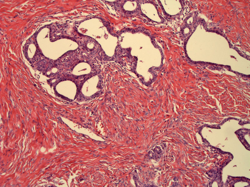
Variably-shaped glands are embedded in dense ovarian stroma.
)
The glands are lined by endometrial-type epithelium. The columnar cells have amphophilic cytoplasm and round uniform nuclei that is more or less basally oriented.
)
Cytologically, the glands show low grade features of malignancy. Note the prominent nucleoli and mild pleomorphism. There is no infiltrative invasion, and a stromal response is lacking.
)
Psammomatous calcifications may be seen. The glandular cells are crowded, but no more atypical than that seen in complex atypical hyperplasia of endometrium.
)
This gross specimen correlates with the previous microscopic images. Borderline endometrioid tumors are typically solid with a tan-white cut surface
)
Endometrioid adenocarcinoma is the second most common type of surface epithelial tumors of the ovary, following serous tumors. Like their counterpart in the endometrium, the range of histologic changes include simple and complex hyperplasia with or without atypia. These tumors arise from the surface epithelium of the ovary, either directly or via transformation of a benign endometrioid adenofibroma. Malignant transformation of endometriosis is also another pathway of oncogenesis.1,2
Benign endometrioid adenofibromas are rare and account for ~10% of all ovarian adenofibromas. Cystic or tubular glands lined by a single layer of endometrial-type columnar cells are surrounded by fibrous stroma. The epithelium may be pseudostratified similar to proliferative-phase endometrial glands, but unlike true proliferative endometrial glands (in the endometrium), mitotic activity is usually absent.
Borderline endometrioid tumors include a group of tumors with designations such as adenofibromas with epithelial atypia, proliferating endometrioid tumors, endometrioid adenofibromas with low malignant potential. The WHO currently groups all these tumors under the umbrella term 'borderline endometrioid tumors'.1
In low-grade borderline tumors, the glandular component may exhibit increased glandular crowding with foci of cribriforming. There may be mild to moderate nuclear atypia and pseudostratification. In the moderate or high-grade borderline tumors, the glands become increasingly crowded with complex architecture. The cytologic atypia may be quite pronounced and mitotic activity is increased. Note that this is similar to the neoplastic sequence seen in endometrial lesions (simple hyperplasia to complex hyperplasia to complex atypical hyperplasia).
When there is stromal invasion or confluent glandular growth, the diagnosis changes from borderline to frank carcinoma.
Most benign and borderline endometrioid tumors are unilateral. They are typically solid tumors with a tan-white cut surface. Some higher-grade tumors may have foci of cystic change with papillary excrescences on the cyst wall.
• Ovary : Endometrioid Adenocarcinoma
• Ovary : Endometrioid Adenocarcinoma, Sertoliform Pattern
1 Fletcher CDM, ed. Diagnostic Histopathology of Tumors. 3rd Ed. Philadelphia, PA: Elsevier; 2007: 579-580.
2 Mills SE, ed. Sternberg's Diagnostic Surgical Pathology.4th Ed. Philadelphia, PA: Lippincott Williams & Wilkins; 2004: 2559-2562.