
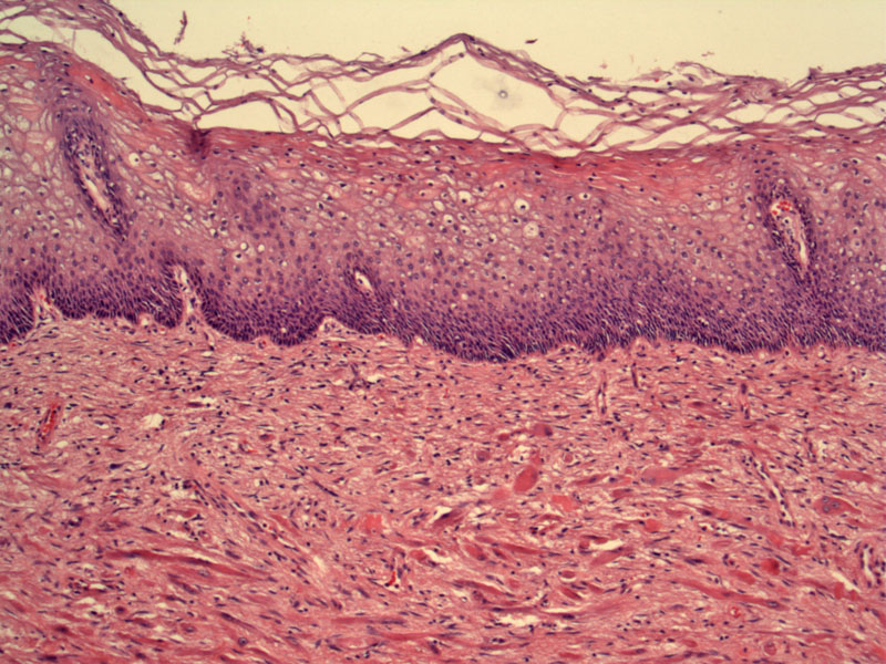
Loosely arranged deeply eosinophilic spindled rhabdomyoblasts are seen in the submucosa. The overlying squamous mucosa is completely normal.
)
The rhabdomyoblasts appear oval and almost inclusion-like when seen en face.
)
Haphazardly placed wavy spindled cells (rhabomyoblasts) are seen. Mitoses are not present. Dilated dilated thin walled capillaries are also present.
)
Note the cross-striations (arrows) in the rhabdomyoblasts.
 are seen in the rhabdomyoblasts. ', 'micro', ''))
Rhabdomyomas are mesenchymal tumors showing skeletal muscle differentiation and occur as either cardiac or extracardiac types. The extracardiac type is classified further into 3 subgroups - adult, fetal and genital.
In the female genital tract, rhabdomyomas are most commonly arise in the vagina, followed by the cervix and vulva. Microscopically, spindled or strap-shaped rhabdomyoblasts with cross-striations are loosely arranged in the loose connective tissue. The vesicular nuclei have a single prominent nucleoli1
The main differential diagnosis is rhabdomyosarcoma botryoides, which occurs in a much younger population and is characterized by a cambium layer and prominent cytologic atypia.
Described in those 25-54 years of age. Presents as a polypoid mass, vaginal bleeding or dyspareunia.
Excision is curative.
Benign; excision is curative. Recurrences are rare and metastases do not occur.
1 Nucci MR, Oliva Esther. Gynecologic Pathology: Foundations in Diagnostic Pathology. Philadelphia, PA: Elsevier: 2009: 111-3.
2 Fletcher CDM, ed. Diagnostic Histopathology of Tumors. 3rd Ed. Philadelphia, PA: Elsevier; 2007: 723.