
Case 1: A cluster of dilated glands filled with mucin is seen. Note the bland attenuated epithelial lining.
)
Case 2: At low power, the cluster of glands can be appreciated.
)
The glands are filled with mucin and the lining is attenuated.
)
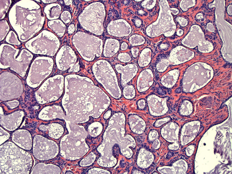
Case 3: The tunnel clusters are well-circumscribed.
)
The lumina can be filled with eosinophilic secretions or mucin (as seen above).
)
The lining of the glands may be distorted, but the cytologic features are benign.
)
Tunnel clusters result from a proliferation of endocervical glands that have side channels emanating from them. They are two main types: (1) dilated glands with a scalloped contour filled with mucin and (2) multiple small acini and tubules lined by columnar or cuboidal cells. The latter type may occasionally exhibit mild focal atypia and mitotic activity and thus, raising the specter of adenocarcinoma. However, on low power, these clusters of dilated glands have a lobular configuration and are well-demarcated. On higher power, the lining epithelium is mostly attenuated and very bland cytologically.
Completely benign finding.
1 Mills SE, ed. Sternberg's Diagnostic Surgical Pathology.4th Ed. Philadelphia, PA: Lippincott Williams & Wilkins; 2004: 2404.
2 Rosai, J. Rosai and Ackerman's Surgical Pathology. 9th Ed. Philadelphia, PA: Elsevier; 2004: 1527.