
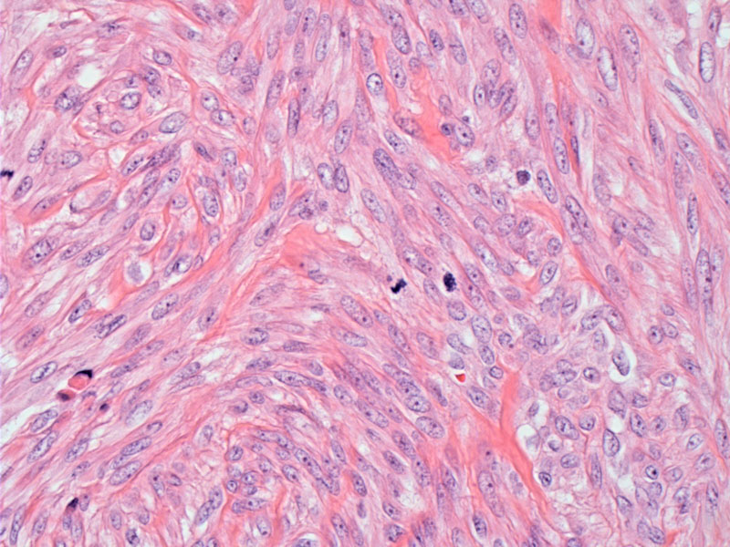
A fasicular growth of spindle cells with blunted bland nuclei without atypia or hyperchromasia. Note the presence of two mitotic figures (center and far left). In studies of these tumors, most of the mitotic counts ranged from 5-9 mitotic figures per 10 high-power fields.
)
This lesion is otherwise a typical leiomyoma (bland cellularity and lack of atypia) but for increased mitotic activity. Two mitotic figures are also seen in this field.
)
This other example showed about 4 mitotic figures per 10 hpf without any atypical forms.
)
Immunostaining for bcl-2 showed diffuse strong positivity. Bcl-2 has been shown to be expressed more frequently and more strongly in leiomyomas compared with leiomyosarcomas and those classified as smooth muscle tumors with unknown malignant potential (Bodner).
)
Except for high mitotic activity ranging from 5-20 mf/10 hpf, this entity exhibits the usual features of leiomyomas such as bland cytology, circumscribed margins and absence of coagulative necrosis (Mills).
According to previous criteria, mitotically active leiomyomas with 5-9 mf/10 hpf have been classified as 'smooth muscle tumors of uncertain malignant potential', and those mitotically active leiomyomas with 10+ mf/10 hpf have been given the diagnosis of leiomyosarcomas. However, it has since been demonstrated that in the absence of cytologic atypia, abnormal mitotic figures or coagulative necrosis, these mitotically active leiomyomas follow a benign course similar to conventional leiomyomas. Care must be taken, however, to thoroughly sample the mass in order to confidently exclude the presence of the aforementioned features of concern (Mills, Fletcher).
The increase in mitotic activity may be due to hormonal influences, as these tumors are found more frequently in pregnancy, during the secretory phase of the menstrual cycle and in patients on hormonal therapy (Fletcher). For example, one study found that patients using progestin preparations had significantly higher mitotic activity in leiomyomas than did control patients (Tiltman).
In effort to distinguish leiomyomas from leiomyosarcomas (LMS) and smooth muscle tumors of uncertain malignant potential (STUMP), Bodner and colleagues studied staining patterns using bcl-2 and p16. They found that leiomyomas are more likely to stain with bc1-2 compared to LMS and STUMP, and in instances where LMS showed strong bcl-2 staining, those patients had less vascular invasion and better survival, suggesting that bcl-2 reactivity may be a good prognostic factor. In another study, Bodner found that LMS and STUMP showed stronger p16 staining compared to leiomyomas. Thus, using bcl-2 and p16 may be helping in resolving between leiomyomas and the more worrisome STUMP or LMS (Bodner, Bodner-Adler).
Same as for leiomyoma.
The long term follow-up of 20 cases (Dgani), as well as patients from other series (Prayson, O'Connor, Bell) supports the view that mitotically active leiomyomas behave in a benign manner. None of the patients in these studies developed recurrence after myomectomy. This data supports the idea that the older terminology 'smooth muscle tumors of uncertain malignant potential' should be discarded.
• Myometrium : Intravascular Leiomyomatosis
• :
• Myometrium : Leiomyoma, Highly Cellular
Bell SW et al. Problematic uterine smooth muscle neoplasms. A clinicopathologic study of 213 cases. Am J Surg Pathol 1994; 18: 535-58.
Bodner-Adler B et al. Expression of p16 protein in patients with uterine smooth muscle tumors: an immunohistochemical analysis. Gynecol Oncol. 2005 Jan;96(1):62-6.
Bodner K et al. Bcl-2 receptor expression in patients with uterine smooth muscle tumors: an immunohistochemical analysis comparing leiomyoma, uterine smooth muscle tumor of uncertain malignant potential, and leiomyosarcoma.J Soc Gynecol Investig. 2004 Apr;11(3):187-91.
Dgani R, et al. Clinical-pathological study of uterine leiomyomas with high mitotic activity. Acta Obstet Gynecol Scand. 1998 Jan;77(1):74-7.
Fletcher CDM, ed. Diagnostic Histopathology of Tumors. 3rd Ed. Philadelphia, PA: Elsevier; 2007: 684.
Mills SE, ed. Sternberg's Diagnostic Surgical Pathology.4th Ed. Philadelphia, PA: Lippincott Williams & Wilkins; 2004: 2516.
O'Connor D, Norris HJ. Mitotically active leiomyomas of the uterus. Hum Pathol 1990; 21: 223-7.
Prayson RA, Hart WR. Mitotically active leiomyomas of the uterus. Am J Clin Pathol 1992; 97: 14-20.
Tiltman AJ. The effect of progestins on the mitotic activity of uterine fibromyomas. Int J Gynecol Pathol 1985; 4: 89-96.