
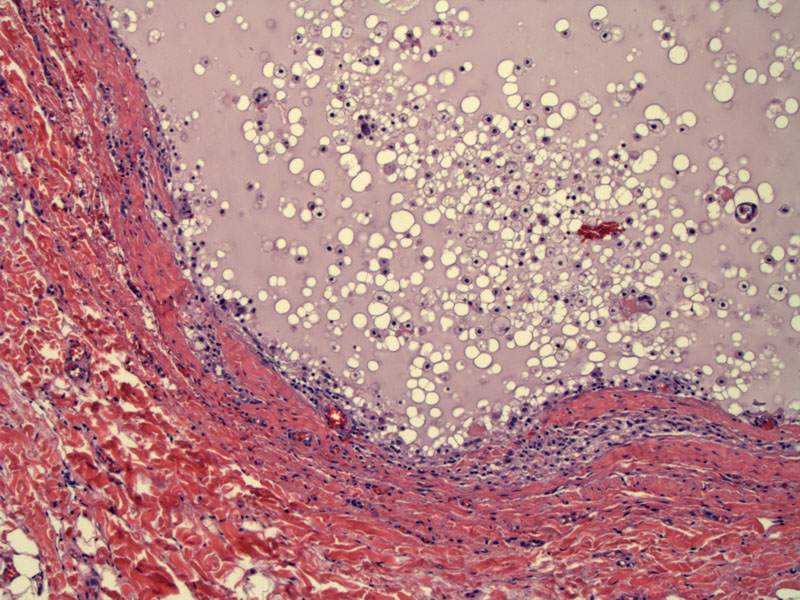
Many cases of thyroglossal duct cyst have lost their epithelium. The lumen is distended by pale blue material with scattered histiocytes
)
Often when intact the lining shows columnar cells, with infoldings of the outer wall.
)
Scattered lymphoid cells may be found in the surrounding stroma.
)
There appears to be cilia lining the luminal aspect of this cyst.
)
If one is lucky, thyroid parenchyma may be found in the wall of the cyst.
)
Cases of papillary carcinoma arising in thyroglossal duct cyst have been the subject of scattered case reports.
Present as a midline mass, which may occur anywhere from the base of the tongue to the thyroid gland; majority are at the level of the thyrohyoid membrane, under the deep cervical fascia. They are midline or just off the midline, and move up and down upon swallowing. They may become secondarily infected.
Excellent. Infected ones may be difficult to excise fully, and as such have a chance of recurrence.