
FNA shows a moderately cellular specimen consisting of uniform slightly spindled cells, which appear relatively bland.
)
The tumor/lung interface shows a well-demarcated tumor.
)
The tumor consists of relatively uniform fat oval cells with a noticeable vascular background and little stroma.
)
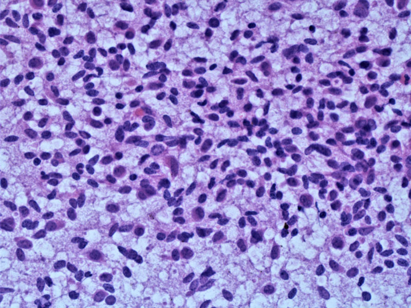
The spindled tumor cells are irregularly arranged. Oval to slightly spindled nuclei are present with a moderate amount of light pink cytoplasm.
)
Focally there is interspersed irregularly distributed collagen within the stroma.
)
Consistent with a carcinoid, the synaptophysin is strongly positive.
)
And the chromogranin is also strongly positive.
)
TTF-1 along the tumor/normal interface shows tumor cell positivity -- TTF-1 has been shown to be positive in about 1/3 of pulmonary carcinoids, and lacking in carcinoids derived from the GI tract and pancreas. (Srivastava)
)
The CDX2 is negative, as expected; in contrast to pulmonary carcinoid, CDX-2 is expressed in 47% of gastrointestinal neuroendocrine tumors as follows: 100% appendiceal, 86% small intestinal, 75% colonic, 18% rectal, and 0% gastric (Lin).
)
Peripheral carcinoid tumors are those carcinoids that arise distal to segmental bronchi.
The typical age is 60 years old. These tumors are not visualized bronchoscopically, usually do not cause symptoms of bronchial obstruction and thus are most often detected incidentally.
Wedge lung resection or lobectomy.
Regional lymph nodes metastases may develop in approximately 1/3 of patients(Abdi). However, despite this, patients usually have an excellent prognosis.
Abdi EA, Goel R, Bishop S, Bain GO. Peripheral carcinoid tumours of the lung: a clinicopathological study. J Surg Oncol. 1988 Nov;39(3):190-6.
Ranchod M, Levine GD.Spindle-cell carcinoid tumors of the lung: a clinicopathologic study of 35 cases. Am J Surg Pathol. 1980 Aug;4(4):315-31.
Srivastava A, Hornick JL. Immunohistochemical staining for CDX-2, PDX-1, NESP-55, and TTF-1 can help distinguish gastrointestinal carcinoid tumors from pancreatic endocrine and pulmonary carcinoid tumors. Am J Surg Pathol. 2009 Apr;33(4):626-32.
Lin X, Saad RS, Luckasevic TM, Silverman JF, Liu Y. Diagnostic value of CDX-2 and TTF-1 expressions in separating metastatic neuroendocrine neoplasms of unknown origin. Appl Immunohistochem Mol Morphol. 2007 Dec;15(4):407-14.