
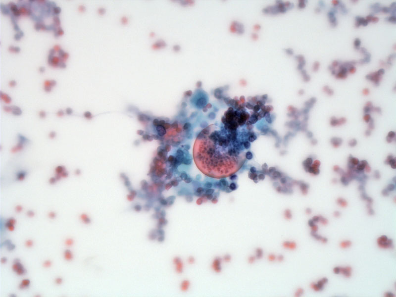
A spherule is seen releasing endospores in a bronchioalveolar lavage (BAL).
)
Another image of a spherule extruding endospores. The spherules are 20 to 60 microns in diameter. In comparison, a red blood cell is approximately 6-8 microns in diameter.
)
Some empty spherules are seen in upper right image.
.', 'cytology', ''))
Center of necrotic pulmonary nodule shows coagulative necrosis and something resembling a spherule.
)
In other areas, degenerating ghost-like spherules can been.
)
Corresponding GMS highlights the degenerating spherule fragments
)
Here is a relatively intact empty spherule -- there are no endosphores inside.
)
Edge of the necrotic pulmonary nodule shows fibrosis, mild chronic inflammation and some multinucleated giant cells occasionally (lower image, just left of center). Relatively normal lung tissue is seen on the right.
)
This case has an admixure of spores including some solid appearing forms.
)
Coccidioides immitis is one of the three main fungal infections that can cause disseminated systemic disease in humans. The other two fungi are Histoplasma capsulatum and Blastomyces dermatitis. These three fungi are dimorphic, meaning at 25 degrees Celsius, they exist as hyphal forms that release spores; and at 37 degrees Celsius (body temperature), they are in yeasts in the form of spherules or ellipses. Whereas Histoplasma and Blastomyces are endemic to the region surrounding the Mississippi River, Coccidioides is endemic to the Southwest (Arizona, New Mexico -- the current residence of yours truly).1,2
These three systemic fungi are acquired through inhalation and may cause granulomatous disease of the lung that mimics tuberculosis. The organisms can also disseminate via the bloodstream to cause systemic disease in immunocompromised hosts.
The clinical symptoms of the three dimorphic fungi can vary. For the majority of individuals, the infection is asymptomic or a mild self-limited respiratory illness. Approximately 10% of patients have lung disease, usually a pneumonia with fever and cough. CXR may reveal pulmonary infiltrates. Granulomas may form with calcifications seen in older lesions. 1% of patients will develop disseminated systemic disease, with involvement of meninges, skin, bones, lymph nodes, liver and spleen.1,2
Coccidioidomycosis with manifestations of pneumonia, arthralgias and skin findings is also known as San Joaquin Valley fever.
Itraconazole or amphotericin B is required to treat chronic or systemic disease.
• Infectious : Coccidioidomycosis
• Small Intestines : Histoplasmosis
1 Kumar V, Abbas AK, Fausto N. Robbins and Cotran Pathologic Basis of Disease. 7th Ed. Philadelphia, PA: Elsevier; 2005: 754-5.
2 Gladwin M, Trattler B. Clinical Microbiology Made Ridiculously Simple. 4th Ed. Miami, FL: MedMaster; 2009: 274-5.