
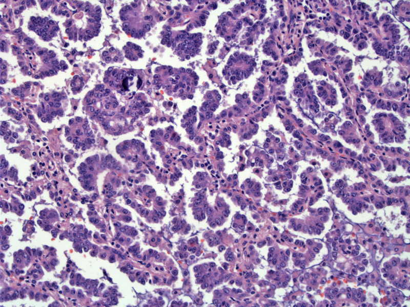
Papillary structures fill the airspaces.
)
Psammoma bodies (calcifications) are abundant in the stroma.
)
A high power view of the psammoma bodies.
)
Papillary structures are admixed with typical glands seen in conventional adenocarcinoma.
)
Papillary structures may be seen in an otherwise conventional pulmonary adenocarcinoma. If this pattern is predominant, the lesion can be diagnosed as "adenocarcinoma, papillary type". Papillae with fibrovascular stalks fill the airspaces and psammoma bodies (similar to those seen in ovarian papillary carcinomas) are common. If the lesion is entirely papillary without recognizable conventional areas, one must consider the diagnosis of metastases from the ovary or thryoid as these organs also produce papillary carcinomas with psammomatous calcifications (Fletcher, Wick).
Several studies illustrate a higher than usual incidence of EGFR mutations in papillary and micropapillary adenocarcinomas of lung (De Oliveira Duarte Achcar)
Papillary adenocarcinomas have significantly worse prognosis compared to bronchioloalveolar carcinomas of the lung.
Leslie KO, Wick MR. Practical Pulmonary Pathology. Philadelphia, PA: Elvesier; 2005: 571.
Fletcher CDM, ed. Diagnostic Histopathology of Tumors. 3rd Ed. Philadelphia, PA: Elsevier; 2007: 184.
De Oliveira Duarte Achcar R, Nikiforova MN, Micropapillary lung adenocarcinoma: EGFR, K-ras, and BRAF mutational profile. Yousem SA. Am J Clin Pathol. 2009 May;131(5):694-700.