
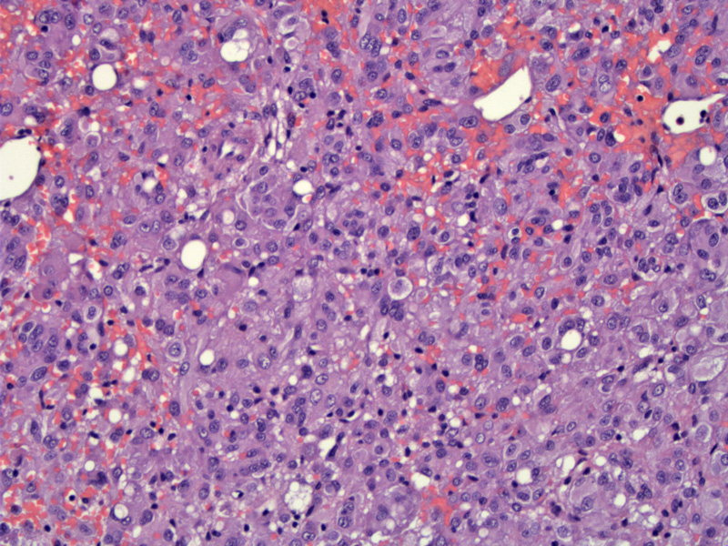
Resembling epithelial cells, these neoplastic cells have a round to polygonal shape with abundant eosinophilic or sometimes clear cytoplasm. There is mild to moderate nuclear atypia here.
)
Rounded to polygonal tumor cells contain abundant eosinophilic cytoplasm. They can grow in diffuse sheets (seen here) or form cords and psuedoglandular spaces.
)
If greater than 50% of the tumor cells in a leiomyoma has an epithelioid appearance (rounded tumor cells with abundant eosinophilic or clear cytoplasm), the tumor is classified as an epithelioid leiomyoma. If the epithelioid leiomyoma demonstrates malignant features (defined below), it becomes a epithelioid leiomyosarcoma. The epithelioid cells are most often arranged in a diffuse sheet, however, nest, cords and pseudoglandular formation can also be seen (Fletcher, Nucci).
Epithelioid leiomyosarcoma is distinguished from its benign counterpart (epithelioid leiomyoma) by the following: (1) significant nuclear atypia; (2) mitotic activity greater than 3 to 4 MF/10 HPF; (3) presence of tumor necrosis (Prayson).
The main entity on the differential diagnosis is a uterine PEComa (perivascular epithelioid cell tumor). In a study of 18 epithelioid smooth muscle tumors of the uterus, all cases (18/18) were negative for CD1a and most (17/18) were negative for HMB-45 (Fadare). In contrast, the literature provides ample evidence that PEComas are CD1a and HMB-45 positive. Thus, these two IHC stains will be helpful in the rare situation that you need to distinguish between the two entities.
Due to the rarity of this tumor subtype, very few studies have been published. However, malignant behavior appears to be associated with increased nuclear atypia, increased mitotic activity, the presence of tumor necrosis and infiltrative borders (Fletcher)
• Myogenic : Leiomyosarcoma, Epithelioid Type
Fadare O, Liang SX. Epithelioid smooth muscle tumors of the uterus do not express CD1a: a potential immunohistochemical adjunct in their distinction from uterine perivascular epithelioid cell tumors. Ann Diagn Pathol. 2008 Dec;12(6):401-5. Epub 2008 Jul 7.
Fletcher CDM, ed. Diagnostic Histopathology of Tumors. 3rd Ed. Philadelphia, PA: Elsevier; 2007: 691.
Nucci MR, Oliva Esther. Gynecologic Pathology: Foundations in Diagnostic Pathology. Philadelphia, PA: Elsevier: 2009; 273-4.
Prayson RA, Goldblum JR, Hart WR. Epithelioid smooth-muscle tumors of the uterus: a clinicopathologic study of 18 patients. Am J Surg Pathol. 1997 Apr;21(4):383-91.