
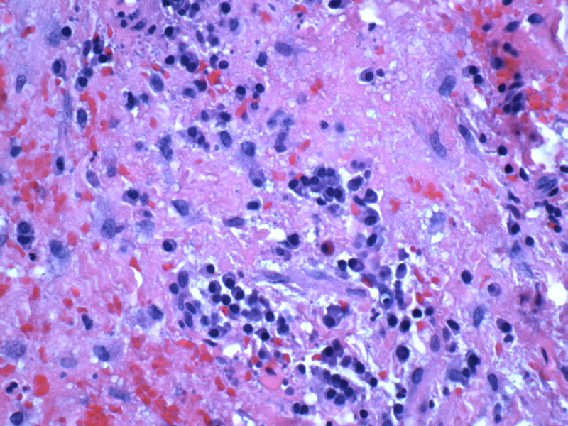
There is a mixed infiltrate consisting of lymphocytes, plasma cells and occasionally histiocytes and neutrophils.
)
This periapical granuloma has a fibrous wall lined by squamous epithelium and thus would be better classified as a periapical cyst.
)
As with a periapical granuloma, granulation tissue with a prominent lymphoplasmacytic infiltrate is present.
)
Periapical granulomas represent 75% of apical inflammatory lesions. It is actually not a true granuloma. This condition describes the mass of inflamed granulation located at the root (apex) of a nonvital tooth, and thus, the term chronic apical periodontitis may be a more accurate description.
Histologically, it consists of granulation tissue with a mixed inflammatory infiltrate. There may be a prominent presence of plasma cells with Russell bodies (eosinophilic globules of gamma globulin). If a true epithelium forms, then the entity may be called a periapical cyst or radicular cyst.
These lesions are usually asymptomatic, but pain and tenderness may occur if there is an acute exacerbation. On imaging, there is a discrete radiolucent area surrounding the root of the tooth.
Removing the infected portions of the tooth pulp via root canal and antibiotics are the key treatment strategies.
Neville BW, et al. Oral & Maxillofacial Pathology 2nd Ed. Philadelphia, PA: WB Saunders; 2002: page 113-115.