
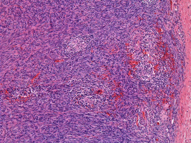
In the ovarian cortex, scattered epithelioid granulomas with associated scant lymphoid cells can be seen.
)
Epithelioid histiocytes that merge with multinucleated giant cells are present in this well-demarcated granuloma -- there is no caseation present.
)
Ovarian granulomas are distinctly uncommon. In developing countries, the most common cause appears to be tuberculosis infections involving the ovaries, fallopian tubes and endometrium. Isolated cases of granulomatous oophoritis from schistosomal infections have also been reported. In developed countries, only a handful of cases are reported, mostly due to noninfectious etiologies.
McCluggage and colleagues1 have conducted the largest study of ovarian granulomas to date and have found 32 cases in their files. The attributed causes of the 32 ovarian granulomas are as follows: foreign body reaction to suture (15), Crohn disease (4), previous diathermy (2), tuberculosis (2), postoperative necrotising reaction (2), endometriosis (1), tubo-ovarian abscess (1) and idiopathic (5).
1 McCluggage WC, Allen DC. Ovarian granulomas: a report of 32 cases. J Clin Pathol 1997; 50:324-7.