
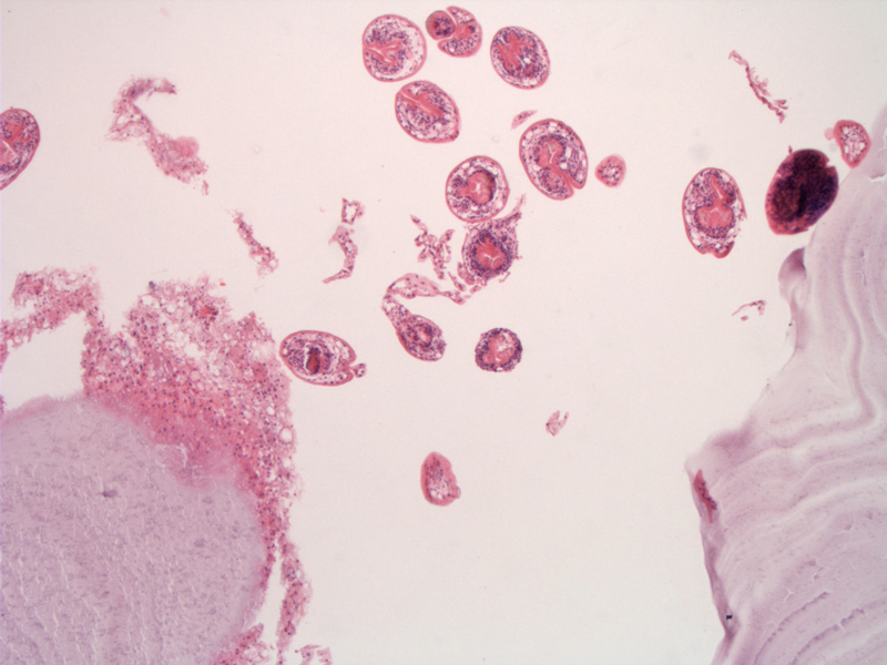
Note the paucity of inflammation but the presence of the brood capsules and membrane. In the intermediate host, once the larvae reach the liver they form a fluid filled hydatid cyst with an inner germinal membrane that produces brood capsules.
)
High power showing brood capsules from which the scolices are seen.
)
The cyst wall shows fibrosis and calcification.
)
Echinococcus granulosus is a cyclophyllid cestode that parasitizes the small intestine. The disease is an emerging disease found in many parts of the world.
Infection is often subclinical, but may manifest with hepatomegaly and ascites. The larveal stage of the parasite usually infects the liver although lung, CNS, and even bone involvement may be present.There is a risk of anaphylaxis if the cyst is aspirated. Secondary complications may arise as a result of infection of the cyst or leakage of the cyst.
The hepatic cysts appear calcified and septated by CT imaging. They can be quite large, and have a tendency to involve the right hepatic lobe.
Excision of the cyst and a course of Albendazole is also recommended.