
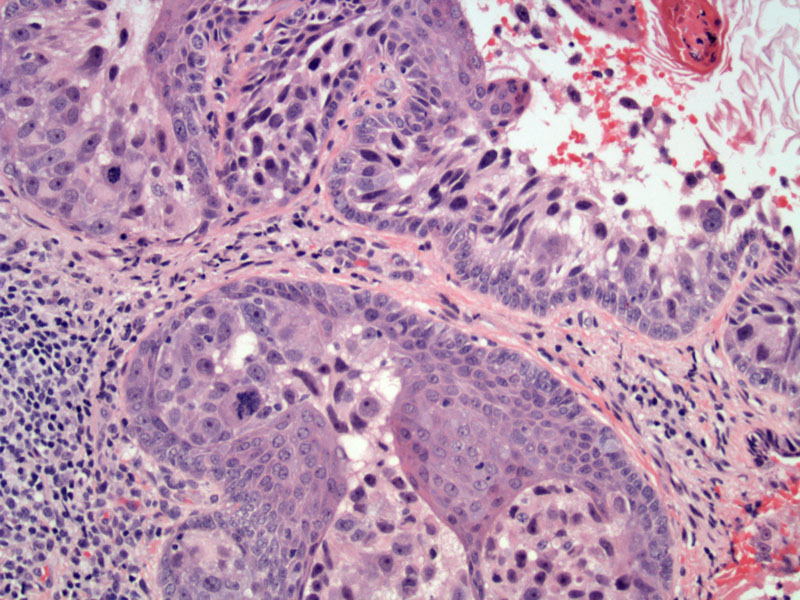
Malignant ductal cells with clear cytoplasm and prominent nucleoli are colonizing the epithelium. The large pleomorphic cells can invade as a single cell or as groups. Note the cluster of malignant cells on the lower portion of this image.
)
Paget's disease of the nipple occurs in ~2% of breast cancers and represents high-grade DCIS extending from a subareolar duct into the squamous epithelium of the nipple. DCIS is always present and an invasive ductal carcinoma is found ~35-50% of the time.1
Classically, the nipple appears as a red, crusted and scaly rash, virtually indistinguishable from an eczematous rash.
Microscopically, one may need to distinguish Paget's disease of the nipple from malignant melanoma or Bowen's disease/squamous cell carcinoma in-situ. This can be difficult as atypical melanocytes and atypical keratinocytes may have similar appearance to atypical ductal cells. Thus, IHC will be helpful. Melanocytes would stain with S100, HMB24 and MelanA; and would be negative for epithelial markers and Her2/Neu. Atypical keratinocytes would stain positively for HMWKs and negatively for Her2/Neu. Finally, 90% of atypical ductal cells are positive for Her2/Neu.1,2
1 Fletcher CDM, ed. Diagnostic Histopathology of Tumors. 3rd Ed. Philadelphia, PA: Elsevier; 2007:928.
2 O'Mallye FP, Pinder SE. Breast Pathology: Foundations in Diagnostic Pathology. Philadelphia, PA: Elvesier; 2006:139-141.