
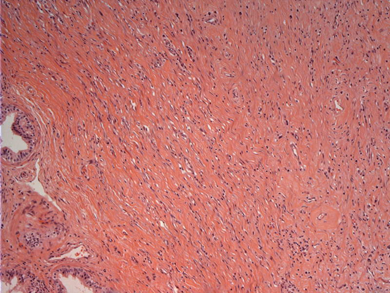
A hypocellular proliferation of spindle cells admixed with hyalinized collagen is seen. On the left, efferent ductules of the epididymis can be seen.
)
Areas of the fibrous lesion can be extensively hyalinized.
)
A concomitant inflammatory component is characteristic. Note the presence of plasma cells between dense sclerotic random collagen bundles. An older term for this entity is inflammatory pseudotumor.
)
Fibrous pseudotumor, also known as fibrous periorchitis, is a reactive non-neoplastic process that mimics a testicular or paratesticular tumor. A diffuse or nodular thickening of the tunica albuginea, epidiymis or spermatic cord is seen. Some experts prefer to call masses and nodules as "fibrous pseudotumor" and reserve the term "fibrous periorchitis" for the diffuse process that encases the testis (Fletcher).
Grossly, singular or multiple nodules are seen adjacent to the testis. The cut surface is firm and white. Microscopically, a proliferation of spindle cells is arranged in whorls and fascicles, often accompanied by inflammatory cells (lymphoctyes, plasma cells, histiocytes and eosinophils). The stroma can be loose or heavily collagenized. Calcifications and ossifications may be seen in degenerate lesions.
It is important to recognize this entity and not misdiagnose it as sarcoma or sarcomatoid mesothelioma, which are more pleomorphic with high mitotic activity.
Seen essentially at any age with a peak incidence in the 3rd decade.
Cheng L, Bostwick DG, eds. Essentials of Anatomic Pathology. 2nd Ed. Totowa, NJ: Humana Press; 2006: 1269.
Fletcher CDM, ed. Diagnostic Histopathology of Tumors. 3rd Ed. Philadelphia, PA: Elsevier; 2007: 846.