
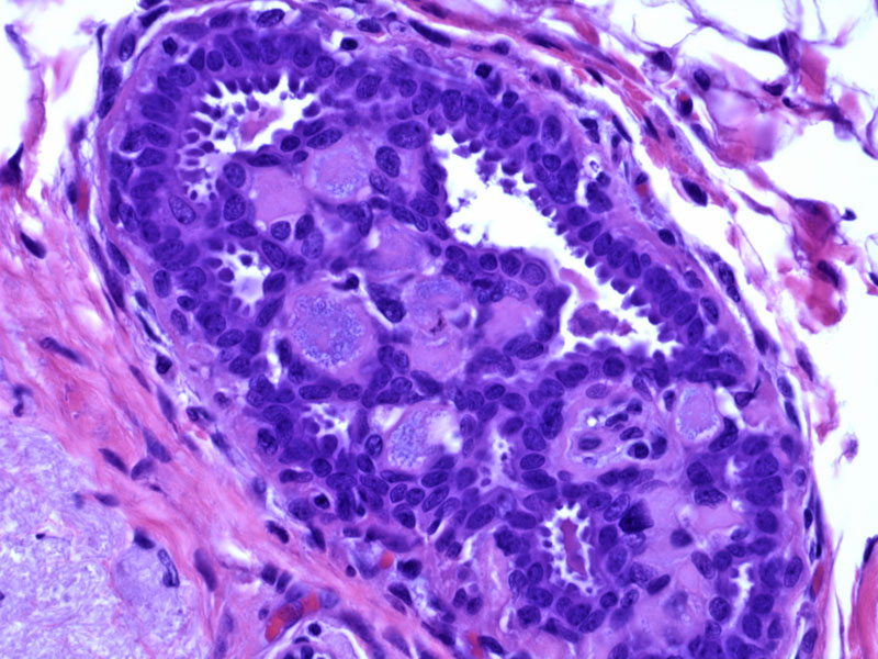
Aggregates of acellular spherules imparts a cylindromatous or cribriform appearance. The surrounding cells are a proliferation of epithelial and myoepithelial cells, although it is the myoepithelial cell that directly surrounds the spherules.
)
Collagenous spherulosis is a form of benign intraductal proliferation with a peculiar morphology. The expanded ducts demonstrate numerous acellular spherules in a background of epithelial and myoepithelial cells. Staining of myoepithelial cells have demonstrated that myoepithelial cells surround each spherule and elaborate a ground substance into these lumens. The spherules may have an eosinophilic, fibrillar, collagneous (hence the term 'collagenous spherulosis) or nearly transparent appearance.
A basophilic appearance of spherules is called mucinous spherulosis, which is thought to be an earlier form of collagenous spherulosis.
Spherulosis is usually an incidental finding in breast biopsies or mastectomies. Also, collagenous spherulosis can become calcified, leading to mammographically detectable microcalcifications. This lesion is often found in conjunction with other proliferative lesions such as papillomas, sclerosing adenosis, radial scars, atypical intraductal hyperplasia and lobular intra-epithelial neoplasia.1,2
The significance of spherulosis is that it may be under-recognized and misdiagnosed as an atypical or malignant entity, namely adenoid cystic carcinoma and DCIS. All these entities may have a cribriform appearance. Note also, that in certain instances, spherulosis may occur in conjunction with LCIS, in which the proliferative epithelial and myoepithelial cells are replaced by neoplastic lobular cells with mildly atypica.
1 Rosen PP. Rosen's Breast Pathology. 3rd Ed. Philadelphia: Lippincott Williams & Wilkins; 2009:130-3.
2 Mooney EE et al. Spherulosis of the Breast: A Spectrum of Mucinous and Collagenous Lesions. Arch Pathol Lab Med. 1999;123:626-630.