
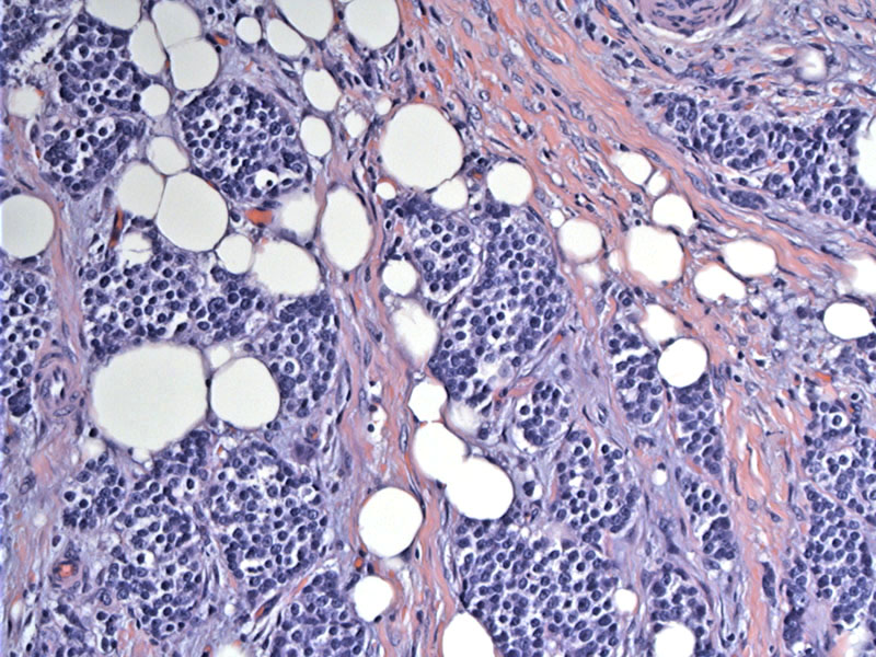
The tumor invades surrounding fat, but retains its nested growth pattern.
Irregular nests of monotonous cells with fibrous septation can be appreciated. The tumor cells exhibit the characteristic finely dispersed chromatin and inconspicuous nucleoli.
Areas of punctate tumor necrosis is consistent with the designation of atypical carcinoid. The tumor was positive for chromogranin.
Primary neuroendocrine tumors of the thymus are thought to arise from neuroendocrine cells that normally reside in the thymus (Fletcher). As with other neuroendocrine tumors, they are subclassified as carcinoid (well differentiated), atypical carcinoid (moderately differentiated), and small cell/large cell neuroendocrine carcinoma (poorly differentiated). These are uncommon tumors and appear to behave more aggressively compared to bronchial carcinoids (Weidner).
Features that characterize atypical carcinoids include increased mitotic activity 2-10 MF per 10 HPF and the presence of coagulative necrosis, which may be punctate(Fletcher).
There is a male predominance (M:F of 3:1). Patients may be asymptomatic, present with symptoms related to a mediastinal mass (couch, chest pain, SVC syndrome). About 1/3 experience Cushing syndrome due to secretion of ectopic ACTH (Weidner, Fletcher).
Up to 25% of thymic carcinoids are associated with MEN type I (Fletcher). Note, however, that thymic neuroendocrine tumors are not associated with the paraneoplastic syndromes seen in thymomas such as myasthenia gravis and hypogammaglobulinemia. Carcinoid syndrome is also not seen in this tumor.
Distant metastases (most commonly to the lung, bone and liver) occurs in a third of the cases. The prognosis is poor and the tumor is generally resistance to chemotherapy or radiation (Fletcher).
- Neuroendocrine carcinomas of the thymus may take the form of either moderately and well-differentiated tumors (thymic carcinoid) or poorly differentiated neoplasms (small cell neuroendocrine carcinoma)
- Unlike neuroendocrine tumors in other sites, those in the thymus are potentially quite morphologically varied -- those with spindle cells, with mucinous stroma, with divergent cell lines including mesenchymal elements and amyloid-like stroma and cases with small cell carcinoma or oncyocytic features have been described (Moran)
Fletcher CDM, ed. Diagnostic Histopathology of Tumors. 3rd Ed. Philadelphia, PA: Elsevier; 2007: 1337-8.
Weidner N, et al. Modern Surgical Pathology Philadelphia, PA: Saunders 2003: 470-1.
Moran CA, Suster S. Primary Neuroendocrine Carcinoma (Thymic Carcinoid) of the Thymus with Prominent Oncocytic Features: A Clinicopathologic Study of 22 Cases. Mod Pathol 2000;13(5):489–494

)
)
)