
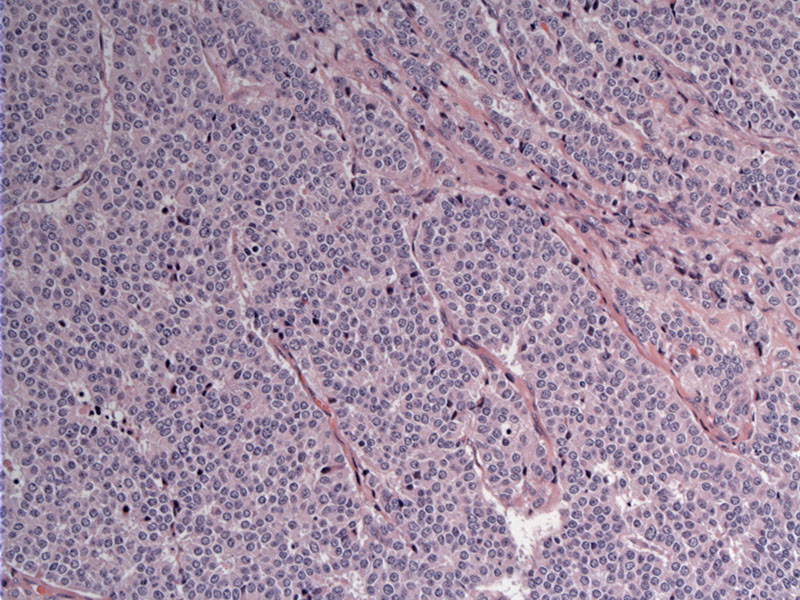
An insular or trabecular growth of round uniform cells, separated by a fibrovascular stroma, is a typical look for a neuroendocrine tumor.
Rosette and ribbon formation can be seen in this well-differentiated NE tumor (carcinoid). The salt and pepper chromatin pattern is obvious.
Thymic neuroendocrine tumors have the same classification as those seen elsewhere i.e. lung or GI tract. Some authors prefer carcinoid , atypical carcinoid, small cell and large cell neuroendocrine carcinomas whereas others prefer well, moderately and poorly differentiated neuroendocrine carcinoma.
Carcinoid tumors of the thymus are rare. In most instances, thymic NE tumors tend to be atypical carcinoids with more aggressive behavior (Rosai). Typical carcinoids have a mitotic count of less than 2 MF per 10 HPF whereas atypical carcinoid have between 2-10 MF per 10 HPF (Fletcher). Up to 25% of these tumors arise in the setting of MEN type I as well as Cushing's syndrome. MEN type I associated tumors tend to be more aggressive, more often occur in men who are smokers (Fletcher).
Mean age is 50 with a male predilection (M:F ratio of 3:1). In a study of 80 thymic neuroendocrine tumors, 1/3 were asymptomatic. The remaining 2/3 of patients experienced chest pain (attributable to a mediastinal mass) and/or endocrine symptoms namely Cushing syndrome (Moran).
Thymic carcinoids tend to be atypical with more aggressive behavior rather than typical and indolent. Encapsulated tumors can be excised, although in many cases, the tumor is invasive with local metastasis to the lymph nodes. The tumor does not respond well to chemotherapy or radiation, thus, 5 year survival is about 30-60% (Fletcher).
Fletcher CDM, ed. Diagnostic Histopathology of Tumors. 3rd Ed. Philadelphia, PA: Elsevier; 2007: 1337-8.
Moran CA, Suster S. Neuroendocrine carcinomas (carcinoid tumor) of the thymus. A clinicopathologic analysis of 80 cases. Am J Clin Pathol. 2000 Jul;114(1):100-10.
Rosai, J. Rosai and Ackerman's Surgical Pathology. 9th Ed. Philadelphia, PA: Elsevier; 2004: 482-3.

)
)