
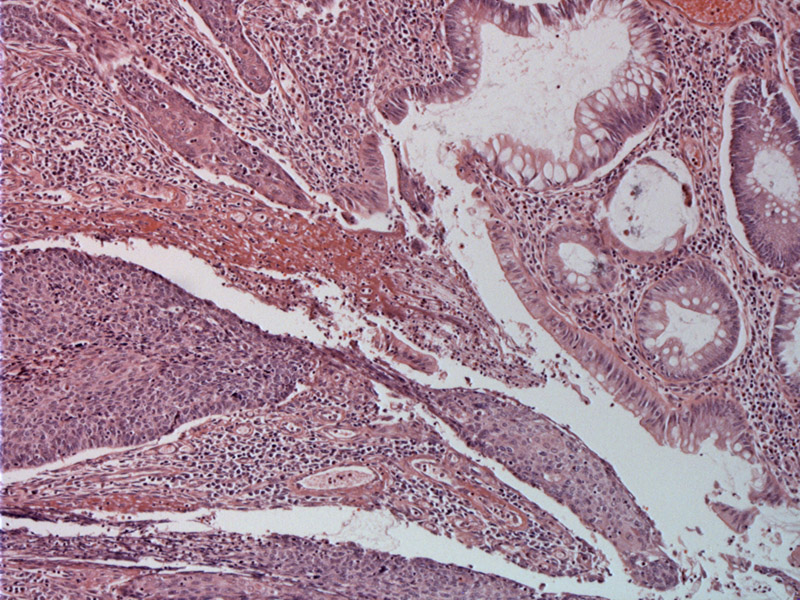
The anorectal junction consists of the transition from simple columnar epithelium with goblet cells to a stratified squamous epithelium. The majority of squamous cell carcinoma of the anal canal arise in this zone. One can see a basaloid proliferation of cells emanating from the surface with a downward growth pattern.
)
The invasive tumor dissects through the stroma and consists of cohesive nests of basaloid cells. There is some evidence of peripheral pallisading.
)
Focal squamoid differentiation with keratinization can often be seen.
)
In the pectinate line of the anal canal, simple columnar epithelium with goblet cells (resembling the colon) abruptly transitions to a stratified squamous epithelium. SCC of the anal canal have two major subtypes: 1. large cell keratinizing or nonkeratinzing, 2. basaloid, also formerly termed cloacogenic carcinoma.
Basaloid carcinoma consists of a diffuse proliferation of small cells (high N/C ratio) with palisading at the periphery of tumor islands. Foci of necrosis or keratinzation can be seen (Fletcher). Large cell and basaloid patterns can be seen within the same tumor, and both subtypes have similar prognosis. The two rarer and more aggressive histologic subtypes of SCC include anaplastic small cell carcinoma (a poorly differentiated form of basaloid SCC) and SCC with mucinous microcysts (Iacobuzio).
Note that this entity must be distinguished from basal cell carcinoma of the perianal skin and neuroendocrine small cell carcinoma.
Risk factors include multiple sexual partners, anal intercourse, smoking, immunosuppression and HIV positivity (Iacobuzio).
Resection (especially for anal margin cancers) with adjuvant chemotherapy or radiation yields a 5 year survival rate of 60% to 89% (Iacobuzio).
Size, depth of invasion and lymph node metastases are important prognostic features (Fletcher).
• Anal : High Grade Squamous Dysplasia (AIN III)
Iacobuzio-Donahue CA, Montgomery EA. Gastrointestinal and Liver Pathology: Foundations in Diagnostic Pathology. Philadelphia, PA: Elsevier; 2005: 411-4.
Fletcher CDM, ed. Diagnostic Histopathology of Tumors. 3rd Ed. Philadelphia, PA: Elsevier; 2007: 409-410.