
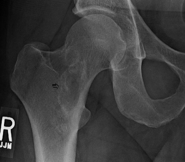
There is a well-circumscribed proximal femur sclerotic lesion seen.
Expansion of the rib is seen by a well circumscribed lesion with a gritty and leather-like consistency. Hemorrhage, cystic change, and foci of blue-gray cartilage may also be present.
Low grade osteosarcoma, the fibrous variant, is a mimicker of fibrous dysplasia. As demonstrated in this image, low grade osteosarcoma (fibrous variant) also shows irregular curvilinear osteoid production with intervening fibrous stroma.
There is a bland fibrous spindle cells proliferation surrounding irregular, curvilinear bony trabeculae, so called Chinese character or hockey stick shapes.
The interface of fibrous and bone tissue shows only minimal osteoblastic rimming. This suggests that osteoblasts are not elaborating bone, rather, that the immature bone is a result of metasplasia of the underlying fibrous tissue.2 The lack of osteoblastic rimming is an important feature to help distinguish this entity from ossifying fibroma (a less common fibro-osseous lesion), in which osteoblastic rimming is present.
Other areas of this osteosarcoma show greater cellularity and mild atypia. Additionally the radiographic appearance of this lesion confirmed aggressive findings.
Fibrous dysplasia is a defect in the maturation of bone. There is growing evidence that the pathogenesis involves an activating missense of the GNASI gene, which encodes a subunit of a stimulatory G protein, is involved (Riminucci, Bianco). Basically the mutation causes the osteoblast to synthesize fibrous tissue in the medullary bone instead of normal bone (Vargas).
Fibrous dysplasia is the most common fibro-osseous lesion and exists in two main forms - the monostotic form (affecting a single bone) and polyostotic form (affecting multiple bones). The most commonly affected bone is the femur, followed by tibia, ribs, maxilla and skull.
There are two syndromes of interest that include the polyostotic form in their presentation. The McCune-Albright syndrome includes the polyostotic fibrous dysplasia, "coast of Maine" cafe-au-lait spots with jagged irregular borders, and endocrinopathies (e.g. precocious puberty). Mazabraud's syndrome refers to polyostotic fibrous dysplasia with multiple intramuscular myxomas.
The classic deformity seen on radiograph is the "shepherd's crook", where the femur is fractured multiple times and heals in a jagged manner resembling a shepherd's crook. In other involved bones, the radiologic images typically show a circumscribed, radiolucent lesion surrounded by a sclerotic rim. The lesional tissue often have a "ground glass" appearance, a finely granular look due to abnormal mineralization (resulting in scattered bone islands).
Histologically, a proliferation of spindle cells in a fibrous stroma in whorls, short fascicles or vague storiform pattern with scattered immature woven bone is characteristic. The bony trabeculae are often described to have a Chinese character or hockey stick appearance. At low power, the trabeculae form C, S or Y shapes. Osteoblastic rimming is minimal.
Osteofibrous dysplasia may present with a similar histologic picture, but in osteofibrous dysplasia, the lesion is intracortical (versus medullary in fibrous dyplasia). Furthermore, there is no osteofibrous dysplasia exhibits osteoblastic rimming of trabeculae.
Fibrous dysplasia tends to present in children and adolescents (peak age between 10-20)(Bullough). There is no clear sex predilection. The most common sites are the proximal femure, ribs, jaw bones and skull.
Focused on relieving deformities. A lesion prone to pathologic fractures can be treated with curettage and bone grafting or pinning in larger lesions involving weight-bearing bones. Smaller lesions in non-weight-bearing bones may be resected.
Although most lesions are asymptomatic and fibrous dysplasia is generally considered a benign lesion, significant morbidity may result certain instances. For example, if fibrous dysplasia involves the craniofacial bones, there may be pronounced facial disfigurement. Some patients will experience multiple pathologic fractures since the abnormally formed fibrous bone is suboptimal for bearing weight.
→Mutations in GNASI (affecting in the G protein pathway) may be involved.
→Involvement of multiple sites by fibrous dyplasia, jagged "Coast of Maine" cafe-au-lait spots, and precocious puberty is called McCune-Albright syndrome.
→Involvement of multiple sites by fibrous dysplasia and multiple intramuscular mxyomas is called Mazabraud's syndrome.
→Histologically, the bony trabeculae form characteristic Chinese character or hockey stick shapes. Osteoblastic rimming is not present.
• Oral Cavity : Ossifying Fibroma
• Fibrous : Myositis Ossificans
Bianco P, Riminucci M, Majolagbe A et al. Mutations of the GNAS1 gene, stromal cell dysfunction, and osteomalacic changes in non-McCune-Albright fibrous dysplasia of bone. J Bone Miner Res. 2000 Jan;15(1):120-8.
Bullough P. Orthopaedic Pathology. 4 Ed. London, UK: Mosby; 2004: 431-7.
Fletcher, Unni, and Mertens. World Health Organization Classification of Tumours: Pathology and Genetics of Tumours of Soft Tissue and Bone. IARC Press: Lyon 2002.
Riminucci M, Liu B, Corsi A et al. The histopathology of fibrous dysplasia of bone in patients with activating mutations of the Gs alpha gene: site-specific patterns and recurrent histological hallmarks. J Pathol. 1999 Jan;187(2):249-58.
Vargas B, Clayer M. Fibrous Dysplasia. eMedicine (Last updated 8/13/08). Available at emedicine.medscape.com/article/1255262-overview

)
)
)
)
)
)