
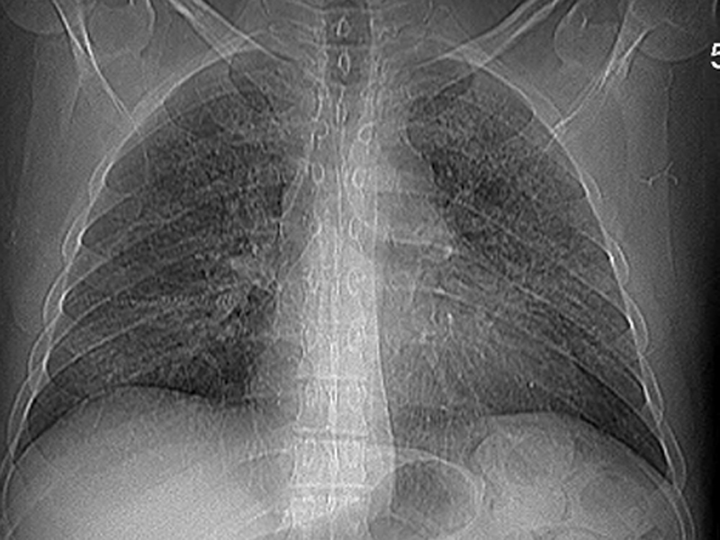
This chest Xray demonstrates the classic non-specific interstitial and ground glass bilateral infiltrate of this infection.
)
Lung parenchyma demonstrates a minimal interstitial chronic inflammation but a marked eosinophilic foamy exudate filling the alveolar spaces.
)
GMS reveals cyst wall structures of the organisms residing within the intra-alveolar exudate. The cysts are round to oval and focally curved or disc shaped.
)
Some of the organisms show central clearing while some may show a targetoid appearance or a cup shape.
)
This bronchioalveolar lavage yielded many casts (arrow). Close inspection shows negative images compatible with Pneumocystis organisms.
)
Numerous organisms are also easily seen on the GMS from this BAL. Note one shows a nice central dot (arrow).
)
Pneumocystis carinii are fungal organisms that affect humans with deficiencies in cell mediated immunity such as those with HIV infection. The species that infects humans is Pneumocystis carinii, and although officially renamed Pneumocystis jiroveci, the new name has yet caught on in all communities.
Pneumocystis carinii is commonly found in the environment and acquired by inhalation. Infection occurs early in life (age 2-3) and 85% of children have been exposed to P. carinii by age 4 based on IgG and IgM levels, and may have had mild respiratory symptoms. This organism remains latent unless the immune system is compromised, in which case, this opportunisitic organism will multiply and cause interstitial pneunomia (Gladwin).
Patients with HIV and low CD4 counts (below 200) are most at risk for infection. Onset may be insidious, with the gradual development of fever, dyspnea, tachypnea and nonproductive cough. Occasional patients may present with a more acute fulminant condition.
Chest X-ray shows a bilateral, diffuse interstitial and alveolar infiltrate, initially in the perihilar and lower lung fields. The early image may show "ground glass" infiltrates, with relative sparing of the upper lungs. However, up to 10-20% of AIDS patients may show no initial X-ray abnormality.
BAL (bronchoalveolar lavage) is high yield for diagnosis. The cells obtained can then be stained using GMS (Gomori methenamine silver) stain to visualize the cysts of P. carinii (Zander).
Treatment includes trimethoprim-sulfamethoxazole (TMP-SMX) and/or pentamidine. Early steroid use may reduce morbidity and mortality. Prophylaxis with TMP-SMX is routine in HIV patients with CD4 counts below 200.
Gladwin M, Trattler B. Clinical Microbiology Made Ridiculously Simple. 4th Ed. Miami, FL: MedMaster; 2009: 330.
Zander DS, Farver CF. Pulmonary Pathology: Foundations in Diagnostic Pathology. Philadelphia, PA: Elvesier; 2008: 770-1.