
Here is a low power image, showing all three zones in cortex and medulla to right.
)
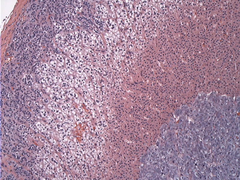
The outermost layers of the adrenal cortex consist of the zona glomerulosa seen to the far right transitioning to zona fasciculata (clear cells). The zona glomerulosa sits immediately beneath the capsule. The zona glomerulosa is the site of aldosterone production.
)
Higher power of zona fasciculata, a broad band of cells that lies between the zona glomerulosa and zona reticularis. The transition between zones is typically indistinct. As seen in this image, the ZF cells have distinct cell membranes, abundant clear cytoplasm, and sometimes a small nucleolus. The cells are filled with lipids such as cholesterol and fatty acids.
)
Another image highlighting the zona glomerulosa beneath the capsule. The cells are often aggregated into small clusters or short cords supported by a small amount of fibrovascular stroma. There is less cytoplasm than in other zones of the cortex. This zone often appears incomplete around the entirety of the gland.
)
This image represents the innermost layer of the adrenal cortex, the zona reticularis. It abuts the adrenal medulla, a small portion of which is seen in the upper left corner. The cells contain granular cytoplasm and are arranged in anastamosing cords.
)
The innermost part of the adrenal is the medulla, which occupies approximately 1/10th the entirety of the organ. The medulla is usually easily recognizable due to its basophilic cells arranged in ill-defined clusters. Mild nuclear variation is a typical feature of this area. A delicate supporting fibrovascular network is also seen.
)
The adrenal glands are located directly above each kidney. A normal adult gland weighs less than 6 grams with a 2 mm thick cortex. The adrenal consists of the inner medulla and the outer cortex.
The outer cortex has three zones and responds to ACTH (adrenocorticotrophic hormone) secreted by the pituitary gland.
- The zona glomerulosa is the outer most zone and produces mineralcorticoids, with aldosterone being the most potent. The function of these steroids is to regulate the body's water and electrolyte balance through the conservation of sodium and elimination of potassium.
- The middle zone is the zona fasciculata, it produces glucocorticoids (glucose + cortex + steroid). These steroids mediate the metabolism of glucose as well as dampen the immune system.
- The innermost zone is the zona reticularis which is known to produce sex steroids, including androgens and estrogens.
The medulla of the adrenal gland is also the body's primary source of epinephrine production and response under stress ("fight or flight"). The medulla contains chromaffin cells, the source of epinephrine and norepinephrine, which are stored in granules.
Eroschenko, Victor P. diFiore's Atlas of Histology with Functional Correlations 10th ed, 2005. Lippincott Williams & Wilkins. Baltimore, MD.
Lack, Ernest E. Tumors of the Adrenal Glands and Extraadrenal Paraganglia." AFIP Atlas of Tumor Pathology, 2007. ARP Press. Silver Spring, Maryland. (1-35)
***Author: Romilly Tsinhnahjinnie, BA-MD student, University of New Mexico. Co-author: Lisa Cerilli MD, Dept of Pathology, University of New Mexico.