
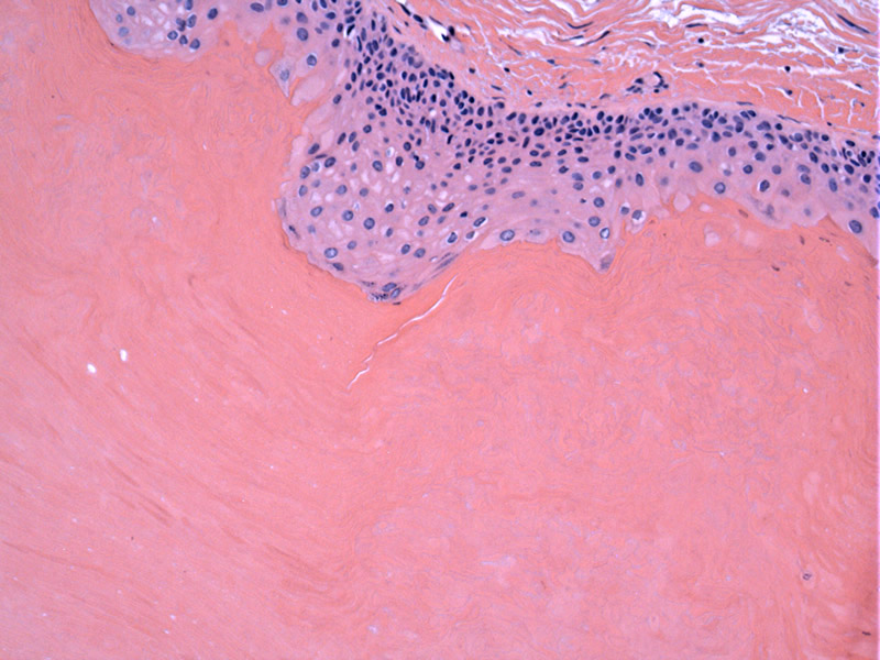
The cysts are filled with keratin and its breakdown products and are lined by walls resembling the external (outer) root sheath of the hair. The squamous epithelium producing the keratin has no granular layer.
)
Calcification of the keratinous material may arise secondarily.
)
Pilar cysts are benign epithelial cysts that are quite common, affecting 5-10% of the population. Most of them (>90%) appear on the scalp.
Also known as trichilemmal cysts, these cysts are derived from the outer root sheath (trichilemma) of the hair follicle. Thus, the keratinization process seen in the cysts is similar to that of the outer root sheath -- rapid keratinization is seen without a granular layer. This is in contrast to an epidermoid cyst where there is a granular layer (Laumann).
Cysts may arise sporadically or in the setting of an autosomal dominant pattern of inheritance. They are most commonly present on the scalp, and there may be multiple cysts.
Excision provides a definitive cure.
Laumann AE. Pilar Cyst (Trichilemmal Cyst): eMedicine. Last updated on November 8th, 2011. Available at: emedicine.medscape.com/article/1058907-overview
Rapini RP. Practical Dermatopathology. Philadelphia, PA: Elsevier; 2005: 253-4.