
Author: Romilly Tsinhnahjinnie, BA-MD student, University of New Mexico.
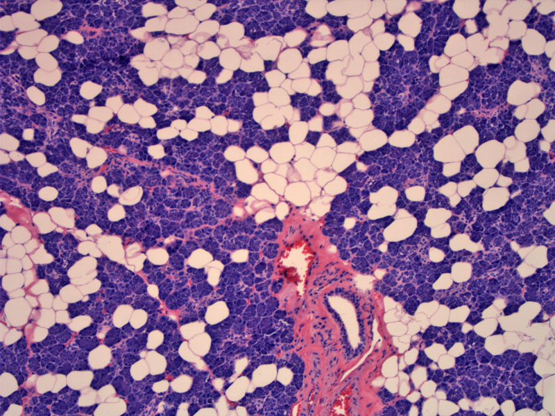
Normal parotid gland is visualized on low power. The acini of the parotid gland are almost entirely serous in type. Intralobular fat is very common.
)
Serous acini are arranged in small spherical clusters of 3-6 cells; numerous zymogen granules impart a deeply basophilic appearance; the ducts in the middle of the image are striated ducts. Cells are slightly columnar with eosinophilic cytoplasm. Image
)
Large interlobular excretory duct in the parotid is seen in the center.
)
Normal parotid gland with acini on FNA (Pap stain).Note the retention of the acinar architecture.
)
The submandibular gland exhibits both mucous (pale staining) and serous cells (dark staining).
)
High power of submandibular gland -- the serous cells are arranged in a way such that they lie in the periphery of the mucous cells, forming demilunes (half moons). The mucous cells contain abundant clear cytoplasm.
)
The normal sublingual gland consists of a mixture of serous and mucous acini. The mucous acini predominate in this gland. Note the numerous mucous cells which have pale staining abundant cytoplasm.
)
The three major glands (parotid, submandibular, and sublingual) convey their secretions (saliva and enzymes) to the oral cavity via excretory ducts. These glands produce approximately 1 liter of saliva per day. Saliva is a made up of water, ions, mucus, electrolytes, antibodies, and digestive enzymes (e.g. amylase, lysozyme). Amylase and lysozyme are produced by serous acini and initiate the breakdown of starch and control bacterial flora, respectively. As we all know, the sight, smell, thought, taste, or presence of food creates the autonomic stimulation of the salivary glands.
There are two major cells that make up the salivary glands: serous and mucous. The serous cells are pyramidal with round nuclei -- granules are present in the apical portion of the cells. Mucous cells are similar in shape, but the cytoplasm is filled with mucus and pale staining. In mixed acini, serous cells form a crescent cap "serous demilune" over the mucous cells.
Surrounding both the serous and mucous acini are the myoepithelial cells, sometimes called basket cells (they surround the acini and form branches resembling a weaved basket). These flattened cells are contractile, propelling the secretory products into ducts. Myoepithelial cells are located between the cell membrane of the secretory cells in acini and the surrounding basement membrane. Often, they can be hard to detect and usually, only the nuclei can be seen.
The parotid gland is the largest salivary gland and weighs 15-30 grams. It is located anterior and inferior to the ear. This gland is purely serous and divided into numerous lobules, each containing many secretory units. The submandibular gland is the second largest gland, weighing 7-15 grams. Located on the floor of the mouth, the gland consists of both mucous and serous acini, usually the serous portion exists as demilunes. The sublingual gland is the smallest of the three glands with a weight of 2-4 grams. It is located inferior to the tongue and is also a mixed gland, with mucous acini predominating.
Serous, mucous, and mixed acini empty secretions into the intercalated ducts. The intercalated ducts are the smallest and merge to form the larger striated ducts. These ducts are lined by tall, columnar cells with large nuclei and basal striations. Striated ducts join to form larger excretory ducts. These ducts then form interlobular and interlobar ducts, the terminal portions of these ducts secrete saliva into the mouth.
Ellis, Gary L. and Auclair, Paul L. "Tumors of the Salivary Glands", AFIP Atlas of Tumor Pathology, 4th series. American Registry of Pathology, 2008. Washington DC. (1-20).
Eroschenko, Victor P. "diFiore's Atlas of Histology with Functional Correlations" 1oth ed, 2005. Lippincott Williams & Wilkins. Baltimore, MD.(284-286).
Klatt, Edward C. "Robbins and Cotran Atlas of Pathology" 1st ed. 2006, Saunders Inc. Philadelphia, PA. (222).
Young, Barbara and Heath, John W. "Wheater's Functional Histology a text and colour atlas" 4th ed. 2000, Harcourt Publishers Limited. Harcourt Place, London (283-284).