
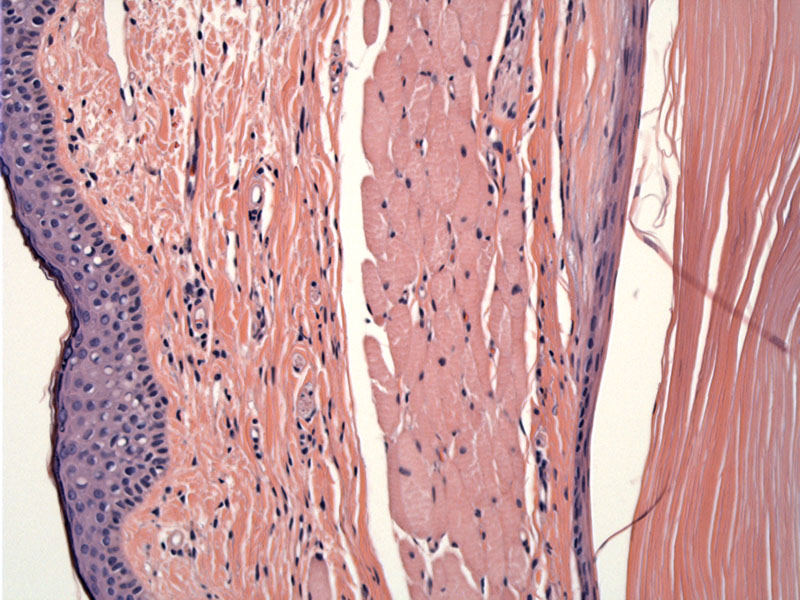
Essentially, this is an epidermal inclusion cyst. Dermal derivative structures (e.g. the pilosebaceous unit) may also be present. A lining composed of squamous epithelium is seen on the right, and the lumen is filled with flakes of keratin. On the left is a thin layer of epidermis with skeletal muscle in between. Cyst rupture can elicit an intense inflammatory reaction.
)
Orbital dermoids (epithelial inclusion cysts) are benign choristomas, choristomas are tumors composed of normal tissue, but in an abnormal location. As the skull sutures lines closed, ectodermal elements were probably pinched off, forming cysts. Approximately 50% of dermoid cysts of the head are located either in the orbit or adjacent to the orbit (Talmadge). Of those in the orbit, the majority (>80%) arise at the superotemporal orbit, at the fronto-zygomatic suture (Cavazza).
Most often seen in children, but can occur at any age. Orbital cysts can expand and cause proptosis. It appears as an egg-shaped soft mass under the skin near the orbital bones. Rupture of the cyst can lead to a severe inflammatory reaction in surrounding tissues.
Complete surgical excision without rupture of the cyst is the ideal treatment (Ahuja).
Ahuja R, Azar NF. Orbital dermoids in children. Semin Ophthalmol. 2006 Jul-Sep;21(3):207-11.
Cavazza S, et al. Orbital dermoid cyst of childhood: clinical pathologic findings, classification and management. Int Ophthalmol. 2011 Apr;31(2):93-7. Epub 2011 Jan 26.
Talmadge C. Orbital Dermoids: eMedicine. Last updated May 28 2010. Available at: emedicine.medscape.com/article/1218740