
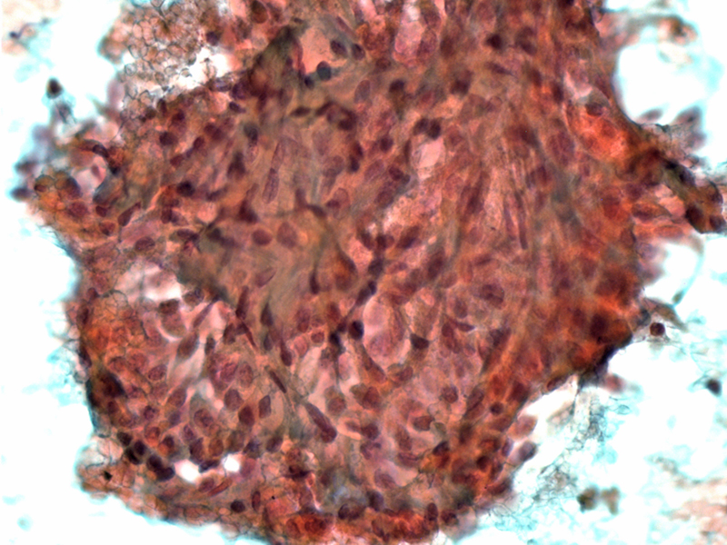
FNA shows a bland uniform cellular epithelial proliferation.
)
The neoplastic myoepithelial cells form vague anastamosing cords and produce abundant basement membrane material. The morphology here is a epitheliod or perhaps plasmacytoid.
)
The nuclei are vesicular and monotonous -- mitotic activity is absent. Atypia, mitoses and necrosis should elicit concern for malignant progression.
)
Myoepithelioma is a benign tumor composed predominantly of myoepithelial cells. The ductal component should be less than 5-10% of the tumor (Fletcher).
Fletcher delineates a most helpful conceptualization of pleomorphic adenoma, basal cell adenoma and myoepithelioma. He views these three neoplasms as entities along a continuum with varying compositions of the two cell types, luminal (acinar or ductal) and abluminal (myoepithelial and basal) cells. On one end, myoepitheliomas are basically "basal cell adenomas minus the ductal component" and basal cell adenomas are "pleomorphic adenomas without the characteristic stroma". Pleomorphic adenomas lie in the middle of the continuum and exhibit both cells types along with its "characteristic strom", which consists of abluminal myoepithelial cells melding into a chondromyxoid stroma (Fletcher).
Myoepithelioma is most commonly seen in the parotid gland, followed by the palate. Grossly, it is a well circumscribed thinly encapsulated tumor with a tan or yellow cut surface.
Histologically, the neoplastic myoepithelial cells have a wide range of morphologies including spindled, plasmacytoid (with abundant eosinophlic cytoplasm and eccentric nuclei), epithelioid, clear or oncocytic. Spindled and plasmacytoid cell types are the most common. The neoplastic cells form sheets, nests trabeculae or fascicles. The stroma can be minimal or abundant with a loose myxoid or dense hyaline appearance (Brandwein, Fletcher).
The malignant counterpart, myoepithelial carcinoma, exhibits cytologic atypia, brisk mitotic activity and infiltrative growth.
Presents as a painless mass and peak age at diagnosis is in the 3-5th decades with equal gender distribution (Fletcher).
Surgical excision is curative.
Brandwein-Gensler M. Head and Neck: Illustrated Surgical Pathology Series. New York, NY: Cambridge University Press; 2010: 311-314.
Fletcher CDM, ed. Diagnostic Histopathology of Tumors. 3rd Ed. Philadelphia, PA: Elsevier; 2007: 258-260.