
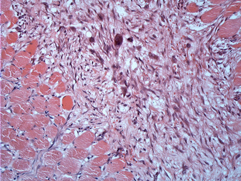
In proliferative myositis, the reactive cellular proliferation extends between and separates muscle fibers, lending a characteristic checkerboard pattern. The individual round muscle fibers appear individual set against the surrounding myofibroblastic proliferation. Note that the muscle fibers are preserved, which helps distinguish this entity from sarcomas and myositis ossificans, where muscle fibers are compromised. Similar to myositis ossificans, 10-20% of proliferative myositis exhibit metaplastic foci of cartilege and bone (Fletcher).
)
The lesion is identical to proliferative fasciitis, composed of a background of (myo)fibroblasts in a loose mxyoid stroma and the scattered presence of ganglion-like cells. These enlarged polygonal cells with basophilic cytoplasm, one or two nuclei with prominent nucleoli are the hallmark features of both proliferative fasciitis and proliferative myositis.
)
There is a reactive look to the ganglion-like or rhabdomyoblast-like cells. There is vesicular chromatin, with distinct but nice round nucleoli. The skeletal muscle is preserved and not compromised as would be seen in a sarcoma. The cells expand the epimysium, perimysium, and endomysium, separating muscle fibers.
)
Proliferative myositis can be considered the intramuscular counterpart of proliferation fasciitis. Due to their deep-seated location, PM can be confused clinically with a sarcoma.
It tends to arise from the skeletal muscles of the shoulder, upper trunk and thigh.
Benign; recurrence is unlikely.
→Considered the intramuscular counterpart of proliferative fasciitis.
→Characteristic "checkerboard" pattern of growth with reactive fibroblastic stroma separating individual muscle fibers.
• Fibrous : Proliferative Fasciitis
Fletcher CDM, ed. Diagnostic Histopathology of Tumors. 3rd Ed. Philadelphia, PA: Elsevier; 2007: 1540-1.
Folpe AL, Inwards CY. Bone and Soft Tissue Pathology: Foundations in Diagnostic Pathology Philadelphia, PA: Elsevier; 2010: 43-5.
Rosai, J. Rosai and Ackerman's Surgical Pathology. 9th Ed. Philadelphia, PA: Elsevier; 2004: 2246.
**Materials courtesy of Dr. Lida Crooks, VAMC Albuquerque NM.