
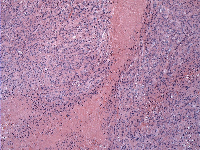
Geographic necrosis is extensive in this case.
)
A sheet-like growth of epithelioid cells can be seen here.
)
Macronucleoli and pleomorphism are evident. Some cells have distinct cell borders with abundant pink cytoplasm.
)
Mitotic activity is usually greater than 20 mf/10 hpf -- There are several two mitotic figures in this field.
)
Yet another high power view demonstrating malignant features (pleomorphism, macronucleoli and sheet-like growth).
)
This is the dura-tumor interface.
)
Irregular infiltration into dura is seen.
)
Ki67 labeling demonstrates the high mitotic index.
)
There was focal limited EMA reactivity, as expected for a dedifferentiated meningothelial tumor.
)
Anaplastic meningioma is similar to atypical meningioma, but with even more malignant features. The diagnostic criteria includes a brisk mitotic rate (>20 MF per 10 HPF) as well as marked nuclear and cellular pleomorphism. All the features that define an atypical meningioma (e.g. necrosis, sheet-like growth with loss of whorls or fasicles, brain invasion, hypercellularity, macronucleoli and brain invasion) are also present.
A poorly differentiated meningioma may appear similar to a high-grade sarcoma, carcinoma or melanoma. Hopefully, the tumor focally retains some features indicating a meningothelial cell origin such as psammoma bodies, however, IHC will probably be necessary to sort through the differential (Prayson).
Keratin staining will be patchy in meningiomas, but diffusely positive in carcinomas. Melanomas will be positive for MelanA and HMB 45, but should be negative in meningiomas; S-100 is variably positive in meningiomas. Unfortunately, mesenchymal markers such as vimentin and EMA are positive in most meningiomas and will not be helpful in distinguishing between high grade sarcomas and anaplastic meningiomas (Prayson).
Along with papillary and rhabdoid variants, anaplastic meningiomas are categorized as WHO Grade III tumors, with median survivals of less than 2 years (Cheng).
• Meninges : Meningioma, Chordoid Variant
• Meninges : Atypical Meningioma
Cheng L, Bostwick DG, eds. Essentials of Anatomic Pathology. 2nd Ed. Totowa, NJ: Humana Press; 2006: 385-6.
Fletcher CDM, ed. Diagnostic Histopathology of Tumors. 3rd Ed. Philadelphia, PA: Elsevier; 2007: 1707-1710.
Kumar V, Abbas AK, Fausto N. Robbins and Cotran Pathologic Basis of Disease. 7th Ed. Philadelphia, PA: Elsevier; 2005: 1409-1410
Prayson, RA. Neuropathology: Foundations in Diagnostic Pathology. Philadelphia, PA: Elvesier; 2005: 489-494.
Prayson R, Kleinschmidt-Demasters BK, Cohen ML. Brain Tumors. Consultant Pathology Series New York, NY: Demos Publishing: 2010: 211-2.