
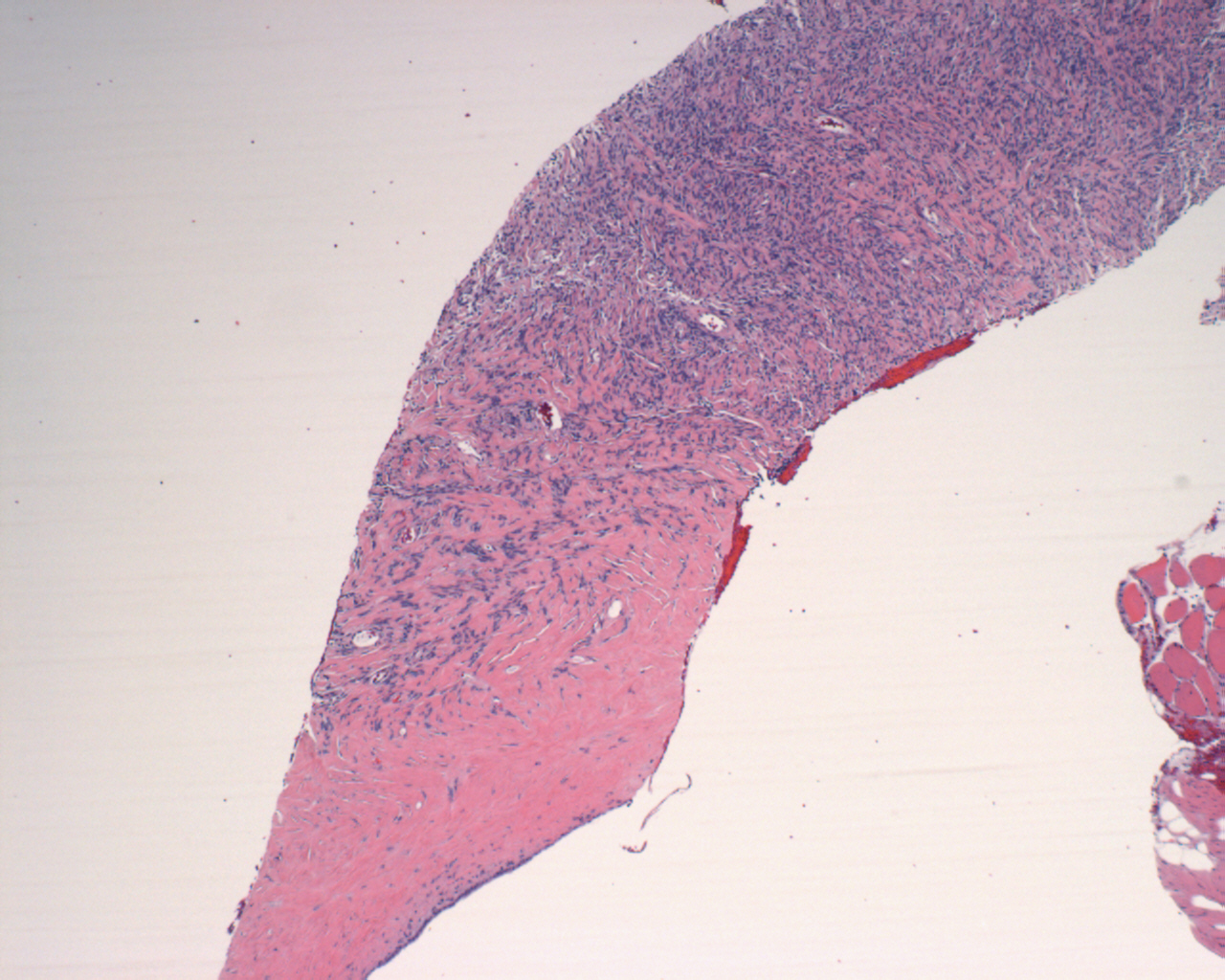
Proliferation of spindle cells and a more sclerotic area.
IMAGE LINKS
 )
 )
 )
 )
 )
CASE BACKGROUND
60 year old female underwent a biopsy for a mass that appeared to be pleural based.
SOLITARY FIBROUS TUMOR
Solitary fibrous tumors can be pleural-based, intraparenchymal (within the lung), or in extrapleural sites such as retroperitoneum, deep soft tissues of proximal extremities, abdominal cavity, trunk, head and neck.Key histologic features include a proliferation of spindle cells with interspersed collagen fibers, an abruption transition from cellular to acellular and sclerotic areas and hemangiopericytoma-like vessels. CD34 and bcl-2 positivity supports this diagnosis. It is prudent to do a cytokeratin stain to exclude sarcomatoid mesothelioma.
RELATED CASES/FURTHER READING
Pleura : Solitary Fibrous TumorFor questions, comments or feedback on this case: editor@surgpath4u.com