
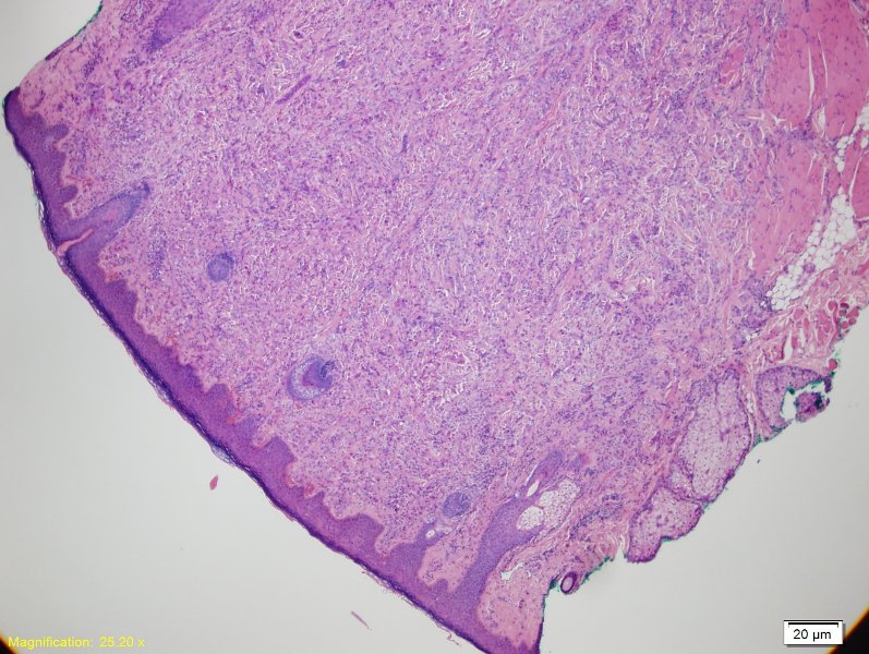
Low power view showing a cellular proliferation filling the dermis
IMAGE LINKS
 )
 )
') )
 )
 )
CASE BACKGROUND
10 year old otherwise healthy female with a small (1.6 cm) cutaneous flesh colored papule on her posterior/medial right arm.
CELLULAR NEUROTHEKEOMA
-This particular case was diagnosed as plexiform fibrohistiocytic tumor (PFHT) by a previous pathologist. Cells were also positive for CD68, SMA, and FXIIIa, and negative for S-100, HMB-45, Melan-A, desmin, and CD34-Cellular neurothekeomas (CNTK) are apart of a larger spectrum of tumors known as nerve sheath myxomas. A less cellular variant with abundant myxoid stroma is known as classic nerve sheath myxoma, while the more cellular variant is known as CNTK
-Benign tumor, but can locally recur
-Cellular variant shows a female preponderance and occurs in a younger population than the classic variant (mean age 24yrs)
-Histologically, basically see a nodular or plexiform pattern of epithelioid to spindled cells infiltrating the dermis and/or subcutis. Cytologic atypia and pleomorphism is usually mild, and the presence of these features do not appear to impact prognosis
-PGP9.5, a broad neural marker, is positive in nearly
RELATED CASES/FURTHER READING
For questions, comments or feedback on this case: editor@surgpath4u.com